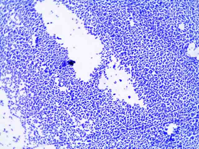
Is S epidermidis Gram positive or negative?
S. epidermidis is a Gram-positive bacterium that appears spherical with an average diameter of 0.5–1.5 µm on light microscopy. The cells of old cultures (>48 h) are often gram-variable to nearly gram-negative. Cells divide in more than one plane to form irregular clusters and aggregates of pairs, tetrads, and short chains.
Is Staphylococcus epidermidis coagulase positive or negative?
Staphylococcus epidermidiswith the highest percentage has the prominent role among coagulase-negative Staphylococci that is the most important reason of clinical infections. Due to various virulence factors and unique features, this microorganism is respected as a common cause of nosocomial infections.
What are the biochemical characteristics of Staphylococcus epidermidis?
The biochemical characteristics of S. epidermidis can be tabulated as follows: S.N Biochemical Characteristics S. epidermidis 1. Capsule Most of the strains are capsulated. 2. Shape Cocci 3. Catalase Positive (+) 4. Oxidase Negative (-) 12 more rows ...
Is S epidermidis a pathogenic disease?
S. epidermidis is mostly non-pathogenic but might cause infections in patients with a compromised immune system. Thus, most of these infections are hospital-acquired. It is a part of the human normal flora as it resides over the epidermis of the skin.
Do Staphylococcus epidermidis produce endospores?
Staphylococci are microbiologically characterized as gram-positive (in young cultures), non-spore-forming, nonmotile, facultative anaerobes (not requiring oxygen). Of significance to humans are various strains of the species S.
Is S. epidermidis positive?
Staphylococcus epidermidis is a coagulase-negative, gram-positive cocci bacteria that form clusters. It is also a catalase-positive and facultative anaerobe.
Is S. epidermidis VP positive or negative?
Biochemical Test and Identification of Staphylococcus epidermidisBasic CharacteristicsProperties (Staphylococcus epidermidis)Nitrate ReductionPositive (+ve)OxidaseNegative (-ve)PigmentNegative (-ve)ShapeCocci42 more rows•Aug 15, 2019
How do you identify S. epidermidis?
As species identification of coagulase-negative staphylococci by fatty acid analyses and biochemical tests is known to be difficult ERIC- and BOX-PCR seem to be excellent tools for the identification of Staphylococcus epidermidis isolates.
How do you distinguish S. aureus and S. epidermidis?
Staphylococcus aureus forms a fairly large yellow colony on rich medium; S. epidermidis has a relatively small white colony. S. aureus is often hemolytic on blood agar; S.
How do you differentiate S. aureus and S. epidermidis?
Furthermore, a significant difference between epidermidis and aureus is that the S. epidermidis colonies are small, round and white in colour while S. aureus colonies are large, smooth and golden in colour. Also, another significant difference between epidermidis and aureus is that the S.
Is Staph epidermidis beta lactamase positive?
Eighty-three per cent of 200 different freshly isolated cultures of Staphylococcus epidermidis produced beta lactamase. Growth in the presence of acridine orange or ethidium bromide or growth at 44 C resulted in a high frequency of loss of the beta lactamase genes in some strains of S. epidermidis.
Is Staphylococcus Gram-positive or negative?
gram-positiveStaphylococcus aureus is a gram-positive, catalase-positive, coagulase-positive cocci in clusters. S. aureus can cause inflammatory diseases, including skin infections, pneumonia, endocarditis, septic arthritis, osteomyelitis, and abscesses.
What are the characteristics of Staphylococcus epidermidis?
S. epidermidis is a very hardy microorganism, consisting of non-motile, Gram-positive cocci, arranged in grape- like clusters. It forms white, raised, cohesive colonies about 1–2 mm in diameter after overnight incubation, and is not haemolytic on blood agar.
What is the empiric treatment for staphylococcus epidermidis?
The choice of empiric therapy for staphylococcus epidermidis infection would be IV vancomycin, as methicillin resistance should be assumed. If the pathogen is methicillin-susceptible, then treatment can be narrowed to beta-lactam antibiotics such as nafcillin and oxacillin.
How does coagulase-negative species survive in harsh environments?
One of the crucial factors allowing coagulase-negative species to survive in a harsh environment is the production of the biofilm. Biofilm formation occurs with initial adhesion to a foreign surface or endothelium, which leads to accumulation into multicellular structures.[4] .
How does staph invade the human body?
Many times, these coagulase-negative staph species invade the human body via prosthetic devices, at which point a small number of microbes travel down the prosthetic device to the bloodstream. The bacteria, then, can produce biofilms that help to protect them from host defense or antimicrobials.[2] .
What causes bloodstream infection in catheters?
Staphylococcus epidermidisand other coagulase-negative staphs are one of the leading causes of catheter-related bloodstream infection. The infection largely occurs as the bacteria migrate from the patient’s skin to the surface of the catheter, but they also can migrate via luminal surfaces.[8] .
What are the symptoms of a catheter infection?
For patients with catheter infection, they may present with localized symptoms such as inflammation, erythema, or purulence around the insertion of the catheter. They also can present with systemic signs such as fever, hypotension, and other signs concerning sepsis. Infectious Endocarditis.
Is Staphylococcus epidermidis a cocci?
Staphylococcus Epidermidis - StatPearls - NCBI Bookshelf. Staphylococcus epidermidis is a coagulase-negative, gram-positive cocci bacteria that form clusters. It is also a catalase-positive and facultative anaerobe. They are the most common coagulase-negative Staphylococcus species that live on the human skin.
Is Staphylococcus epidermidis a gram positive or negative?
Staphylococcus epidermidis is a coagulase-negative, gram-positive cocci bacteria that form clusters. It is also a catalase-positive and facultative anaerobe. They are the most common coagulase-negative Staphylococcus species that live on the human skin.
Why is Staphylococcus epidermidisis a BAI?
Also extracellular polysaccharides production and biofilm formation increase the bacterial stability on different surfaces therefore the antibiotic penetration will be prevented [73].
Which microorganism has the highest percentage of coagulase-negative staph?
Staphylococcus epidermidiswith the highest percentage has the prominent role among coagulase-negative Staphylococci that is the most important reason of clinical infections. Due to various virulence factors and unique features, this microorganism is respected as a common cause of nosocomial infections.
Which cocci produced white colonies on blood agar plates?
Rosenbach in 1884 named the Cocci which produced white colonies on blood agar plates as Staphylococcus albus, thereafter in 1891 Staphylococcus epidermidis albus, in 1908 Albococcus epidermidisand Staphylococcus epidermidisin 1916 were used by Welch et al. [2].
What is the most significant event in clinical microbiology?
One of the most significant events in clinical microbiology is the antimicrobial resistance emergence in nosocomial pathogens. There are many various resistance mechanisms in bacteria; hence some of them may be intrinsically resistant to certain antibiotics or to more than one class of antimicrobial agents.
Can bacteria use glucose?
Although this bacterium can use glucose for growing in anaerobically condition, but producing of coagulase and other agents like mannitol fermentation is negative, while in aerobically situation, the acid production is occurring from different carbohydrates (fructose, maltose, sucrose, and glycerol). Ecology.
What is S. epidermidis associated with?
S. epidermidis is primarily associated with nosocomial infections which are primary associated with permanent medical implant devices like catheters. This bacterium is associated with more than 22% of the bloodstream infections of the central intravenous catheters.
What is the pathogenesis of S. epidermidis?
Infections caused by S. epidermidis have been found to cause invasive infections in selected groups of patients, which include preterm neonates, immunocompromised individuals, and patients with permanent medical devices . The pathogenesis of infection caused by S. epidermidis can be explained as follows:
What is the habitat of Staphylococcus epidermidis?
Habitat of Staphylococcus epidermidis. S. epidermidis is the predominant coagulase-negative Staphylococcus species found in the material of human origin. Humans are the only natural host for this organism. The physiological habitat of S. epidermidis is the skin and mucous membranes of humans and animals.
What is the most frequently isolated species from human epithelia?
What is Staphylococcus epidermidis? Staphylococcus epidermidis is a Gram-positive bacterium belonging to the genus Staphylococcus and is the most frequently isolated species from human epithelia.
What is the classification of a staph?
Classification of species of the genus Staphylococcus is based on various factors like the chemical properties of the cell wall, especially the amino acid composition and sequence of the interpeptide bridges of the peptidoglycan and teichoic acid composition.
What is the habitat of S. epidermidis?
The physiological habitat of S. epidermidis is the skin and mucous membranes of humans and animals. The name ‘epidermidis’ indicates the habitat of the organism. S. epidermidis is the most familiar resident staphylococcal species on human skin in terms of population size.
What is mannitol salt agar?
Mannitol Salt Agar (MSA) Small pink to red colonies are formed on MSA. The media remains red as the bacterium cannot ferment mannitol. This media is a selective media for S. aureus and is commonly used to distinguish S. aureus from S. epidermidis. 3.