What is the role of iodine in the Gram stain process?
What is the role of iodine in the Gram stain process? Iodine is a mordant in the Gram stain technique, which functions to intensify the primary stain. A student creates a Gram stain on a bacterial specimen that has a mix of gram -negative and gram -positive organisms but accidentally forgets the decolorizer step.
Can iodine be added before the primary stain in a Gram stain?
Can iodine be added before the primary stain in a Gram stain? No because the iodine allows the crystal violet stain to bind to the peptidoglycan in cell walls. Why is iodine added after the crystal violet? The heat-fixed cells should look purple at this stage.
Why is iodine used to make a wet mount?
Why iodine is added in wet mount? Lugol’s Iodine is a rapid, non-specific contrast dye that is added to direct wet mounts of fecal material to aid in differentiating parasitic cysts from host white blood cells. Many protozoa and cysts take up the dye and appear brown while other objects in the sample remain clear.
Why is iodine used in starch hydrolysis test?
Why is it an essential laboratory procedure?
- A starch test can be performed on a sample to detect it.
- The iodine testing can be used to help distinguish starch from monosaccharides and disaccharides as well as other polysaccharides.
- The iodine testing is used to distinguish between starch, glucose, and carbohydrate.
- For the detection of hyper- or hypothyroidism, blood iodine testing can be used.
What happens if iodine is not used in Gram staining?
If the iodine is not applied during the Gram stain, then gram positive cells will likely stain pink.
What is the purpose of the iodine in the staining process?
Iodine fixes the crystal violet into the cell wall of the bacteria by working as a mordant. A mordant forms a complex that adheres tightly to the cell...
What is the role of iodine in the Gram stain process quizlet?
Iodine is a mordant in the Gram stain technique, which functions to intensify the primary stain.
How does iodine act as a mordant in a Gram stain?
After applying crystal violet and waiting 60 seconds the excess stain is rinsed off with water. Next, a mordant is used. The mordant is Gram's Iodine. This binds to the crystal violet making a large complex that adheres to the cell membrane.
What is the function of iodine in a gram staining technique?
What Is the Function of Iodine Solution in a Gram-Staining Technique? The iodine solution works as a mordant that makes the stain more more prominent and the crystal violet dye become more tightly bound to the bacterial cell. Adding iodine solution or potassium chloride is the second step in the gram-staining process.
Why are staining agents trapped in Gram positive cells?
The staining agents are trapped within the cells of gram positive bacteria because of its thicker peptidoglycan layer, while gram negative bacteria will be unable to hold on the the stain due to its thin layer. A counterstain is added to the specimen staining it red.
How to stain a grain?
The process of grain staining involves the following steps: Step 1. The bacteria is treated with the crystal violet dye, then the iodine solution. Step 2. After the staining, decolorizing agents are added to the specimen. This dehydrates, shrinks and tightens the peptidoglycan layer of the bacteria. The staining agents are trapped within the cells ...
What is the second step in the gram staining process?
Adding iodine solution or potassium chloride is the second step in the gram-staining process. Gram staining is the common and most widely used technique used to differentiate between gram negative and gram positive bacteria.
What is the color of a counterstain?
A counterstain is added to the specimen staining it red. The red coloration does not change the color of the purple coloration of gram positive cells. Conversely, it adds a red color to gram negative bacteria. ADVERTISEMENT.
How Does Gram Staining Work?
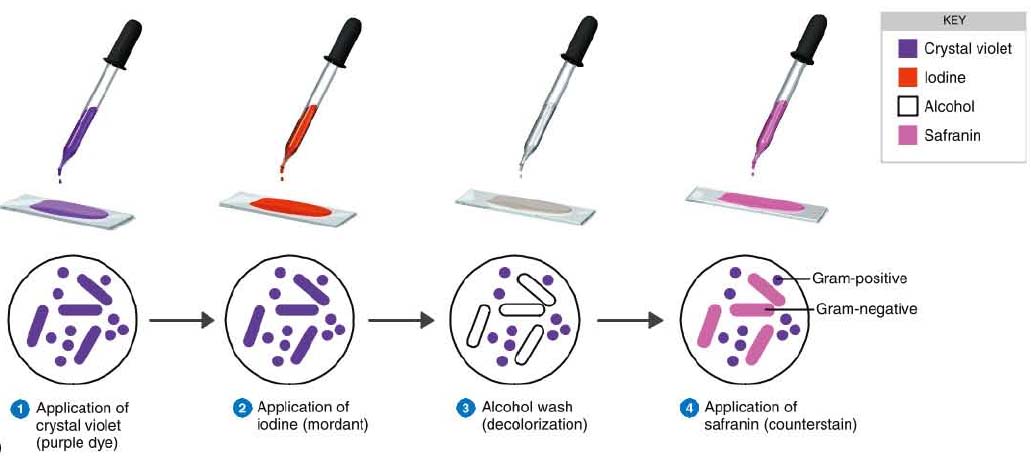
Gram staining involves three processes: staining with a water-soluble dye called crystal violet, decolorization, and counterstaining, usually with safanin. Due to differences in the thickness of a peptidoglycan layer in the cell membrane between Gram positive and Gram negative bacteria, Gram positive bacteria (with a thicker peptidoglycan layer) retain crystal violet stain during the decolorization process, while Gram negative bacteria lose the crystal violet stain and are instead stained by the safranin in the final staining process. The process involves three steps:
Why do Gram positive bacteria stain violet?
Gram positive bacteria stain violet due to the presence of a thick layer of peptidoglycan in their cell walls, which retains the crystal violet these cells are stained with. Alternatively, Gram negative bacteria stain red, which is attributed to a thinner peptidoglycan wall, which does not retain the crystal violet during the decoloring process.
How to stain a cell?
How To- Staining Protocol and Concerns: 1 Make a slide of cell sample to be stained. Heat fix the sample to the slide by carefully passing the slide with a drop or small piece of sample on it through a Bunsen burner three times. 2 Add the primary stain (crystal violet) to the sample/slide and incubate for 1 minute. Rinse slide with a gentle stream of water for a maximum of 5 seconds to remove unbound crystal violet. 3 Add Gram's iodine for 1 minute- this is a mordant, or an agent that fixes the crystal violet to the bacterial cell wall. 4 Rinse sample/slide with acetone or alcohol for ~3 seconds and rinse with a gentle stream of water. The alcohol will decolorize the sample if it is Gram negative, removing the crystal violet. However, if the alcohol remains on the sample for too long, it may also decolorize Gram positive cells. 5 Add the secondary stain, safranin, to the slide and incubate for 1 minute. Wash with a gentle stream of water for a maximum of 5 seconds. If the bacteria is Gram positive, it will retain the primary stain (crystal violet) and not take the secondary stain (safranin), causing it to look violet/purple under a microscope. If the bacteria is Gram negative, it will lose the primary stain and take the secondary stain, causing it to appear red when viewed under a microscope.
What is the process of staining cells with crystal violet?
The process involves three steps: Cells are stained with crystal violet dye. Next, a Gram's iodine solution (iodine and potassium iodide) is added to form a complex between the crystal violet and iodine. This complex is a larger molecule than the original crystal violet stain and iodine and is insoluble in water.
Does alcohol decolorize a cell?
However, if the alcohol remains on the sample for too long, it may also decolorize Gram positive cells. Add the secondary stain, safranin, to the slide and incubate for 1 minute.
What is Gram staining?
The Gram staining is one of the most crucial staining techniques in microbiology. It gets its name from the Danish bacteriologist Hans Christian Gram who first introduced it in 1882, mainly to identify organisms causing pneumonia. Often the first test performed, gram staining involves the use of crystal violet or methylene blue as the primary color. The term for organisms that retain the primary color and appear purple-brown under a microscope is Gram-positive organisms. The organisms that do not take up primary stain appear red under a microscope and are Gram-negative organisms.
What is the first step in gram staining?
The first step in gram staining is the use of crystal violet dye for the slide's initial staining. The next step, also known as fixing the dye, involves using iodine to form crystal violet- iodine complex to prevent easy removal of dye. Subsequently, a decolorizer, often solvent of ethanol and acetone, is used to remove the dye. The basic principle of gram staining involves the ability of the bacterial cell wall to retain the crystal violet dye during solvent treatment. Gram-positive microorganisms have higher peptidoglycan content, whereas gram-negative organisms have higher lipid content.
Do Gram negatives lose their primary stain?
Initially, all bacteria take up crystal violet dye; however, with the use of solvent, the lipid layer from gram-negative organisms is dissolved. With the dissolution of the lipid layer, gram negatives lose the primary stain. In contrast, solvent dehydrates the gram-positive cell walls with the closure of pores preventing diffusion of violet-iodine complex, and thus, bacteria remain stained. The length of decolorization is a critical step in gram staining as prolonged exposure to a decolorizing agent can remove all the stains from both types of bacteria.
How does gram staining work?
Gram staining differentiates bacteria by the chemical and physical properties of their cell walls. Gram-positive cells have a thick layer of peptidoglycan in the cell wall that retains the primary stain, crystal violet. Gram-negative cells have a thinner peptidoglycan layer that allows the crystal violet to wash out on addition of ethanol. They are stained pink or red by the counterstain, commonly safranin or fuchsine. Lugol's iodine solution is always added after addition of crystal violet to strengthen the bonds of the stain with the cell membrane. Gram staining is almost always the first step in the preliminary identification of a bacterial organism. While Gram staining is a valuable diagnostic tool in both clinical and research settings, not all bacteria can be definitively classified by this technique. This gives rise to gram-variable and gram-indeterminate groups.
When to use Gram stain?
Gram stains are performed on body fluid or biopsy when infection is suspected. Gram stains yield results much more quickly than culturing, and are especially important when infection would make an important difference in the patient's treatment and prognosis; examples are cerebrospinal fluid for meningitis and synovial fluid for septic arthritis.
What is the most common gram stain?
A Gram stain of mixed Staphylococcus aureus ( S. aureus ATCC 25923, gram-positive cocci, in purple) and Escherichia coli ( E. coli ATCC 11775, gram-negative bacilli, in red), the most common Gram stain reference bacteria. Gram stain or Gram staining, also called Gram's method, is a method of staining used to distinguish ...
Why is Lugol's iodine added to the gram stain?
Lugol's iodine solution is always added after addition of crystal violet to strengthen the bonds of the stain with the cell membrane. Gram staining is almost always the first step in the preliminary identification of a bacterial organism.
What is the size of a Gram stain of Candida albicans?
Gram stain of Candida albicans from a vaginal swab. The small oval chlamydospores are 2–4 µm in diameter.
What did Gram do to make bacteria more visible?
Gram devised his technique not for the purpose of distinguishing one type of bacterium from another but to make bacteria more visible in stained sections of lung tissue. He published his method in 1884, and included in his short report the observation that the typhus bacillus did not retain the stain.
What is the difference between a purple and a pink gram positive?
Purple-stained gram-positive (left) and pink-stained gram-negative (right) Gram-positive bacteria have a thick mesh-like cell wall made of peptidoglycan (50–90% of cell envelope), and as a result are stained purple by crystal violet, whereas gram-negative bacteria have a thinner layer (10% of cell envelope), so do not retain ...