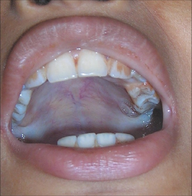
The fauces is a part of the oropharynx directly behind the oral cavity as a subdivision, bounded superiorly by the soft palate, laterally by the palatoglossal and palatopharyngeal arches, and inferiorly by the tongue. The arches form the pillars of the fauces.
What is a faucial pillar?
Faucial pillar. Known as: fauces pillars, palatine arch structure, Palatine arch Expand. The anterior and posterior borders of the tonsillar fossa. They are composed of muscle tissue.
Where is the fauces pillar located in the body?
Faucial pillar. Known as: fauces pillars, palatine arch structure, Palatine arch Expand. The anterior and posterior borders of the tonsillar fossa. They are composed of muscle tissue. Similarly, where is the oropharynx located in the body? Oropharynx.
What is the orientation of the anterior faucial pillar?
anterior faucial pillar. That is, between the soft palate and tongue, it has primarily a vertical orientation. As the anterior faucial pillar blends into the soft palate, the elastic layer assumes a transverse orientation which is just deep to the oral mucous membrane. Theelastic layer was found to have a quan-
What is the faucial pillar of the mouth?
faucial pillar was well-defined in all speci- mens. The bulge which projected medially coursed between the soft palate and tongue (Figure 1). After removing the mucous mem- brane, palatoglossus was found to have a flat- tened belly within the anterior faucial pillar.
What is behind the Faucial pillars?
The fauces is a part of the oropharynx directly behind the oral cavity as a subdivision, bounded superiorly by the soft palate, laterally by the palatoglossal and palatopharyngeal arches, and inferiorly by the tongue. The arches form the pillars of the fauces....Fauces (throat)FaucesFMA55006Anatomical terminology12 more rows
What muscles consist the tonsillar pillars?
The palatoglossus muscle and overlying mucosa form the anterior tonsillar pillar, which is the anterior-most part of the lateral oropharynx. The palatopharyngeal muscle, with its overlying mucosa, forms the posterior tonsillar pillar. The two pillars connect laterally and inferiorly with the lateral oropharyngeal wall.
What are the anterior and posterior Faucial pillars?
Palatoglossal Muscle These are called the anterior and posterior faucial pillars, or the palatoglossal and palatopharyngeal folds, respectively. Beneath the palatoglossal fold is the palatoglossal muscle. It originates from the posterior end of the hard palate and the anterior end of the soft palate.
What nerve Innervates the Faucial pillars?
Sensory events initiating swallowing occur with stimulation to jaw, posterior tongue, faucial pillars, and upper pharynx and are mediated through CN-V, CN-IX, and CN-X.
What is posterior tonsillar pillar?
Posterior tonsil pillar This is the fold of tissue just behind the tonsils. It is created by the palatopharyngeus muscle which extends from the soft palate to the lateral wall of the pharynx.
What separates nasopharynx and oropharynx?
The soft palate divides the pharynx into the nasopharynx dorsally and the oropharynx ventrally. The oropharynx is divided into the isthmus of fauces (throat opening) and a laryngeal part.
What does Faucial mean?
The passage from the back of the mouth to the pharynx, bounded by the soft palate, the base of the tongue, and the palatine arches.
What is Faucial Arch?
(fo'sez) [L.] The constricted opening leading from the oral cavity to the oropharynx. It is bounded by the soft palate, the base of the tongue, and the palatine arches. The anterior pillars of the fauces are known as the glossopalatine arch, and the posterior pillars as the pharyngopalatine arch.
What are anterior pillars?
The anterior pillars are formed by the palato-glossus muscles. The blood supply comes from a branch of the descending palatine artery and is returned by the inferior palatine vein from a plexus surrounding the tonsil, and from the pillars and soft palate into the deep facial vein.
Where is the palatoglossal arch located?
The palatoglossal and palatopharyngeal arches are the two mucosal folds that extend inferiorly from each lateral border of the soft palate. The palatoglossal arch is located anteriorly. It contains the palatoglossus muscle and connects the soft palate with the root of the tongue.
What is the opening to the mouth called?
The pocket-like part of the mouth that is framed on the inside by the gums and teeth, and on the outside by the cheeks and lips is called the oral vestibule. Moving farther into the mouth, the opening between the oral cavity and throat (oropharynx) is called the fauces (like the kitchen “faucet”).
What tongue muscle sticks out your tongue?
the genioglossus muscleThe primary function of the genioglossus muscle is to protrude the tongue anteriorly and deviate the tongue to the opposite side.
What is the anterior pillar of the fauces?
The arches form the pillars of the fauces. The anterior pillar is the palatoglossal arch formed of the palatoglossus muscle. The posterior pillar is the palatopharyngeal arch formed of the palatopharyngeus muscle.
Is faucitis a secondary disease in cats?
Inflammation of the fauces, known as faucitis, is seen in animals. In cats, faucitis is usually a secondary disease to gingivitis but can be a primary disease.
What is the opening at the back of the mouth into the throat called?
(Fauces labelled as Isthmus faucium at center right.) The fauces, isthmus of fauces, or the oropharyngeal isthmus, is the opening at the back of the mouth into the throat. It is a narrow passage between the velum and the base of the tongue.
Which muscles are responsible for the fauces?
The approximation of the arches due to the contraction of the palatoglossal muscles constricts the fauces, and is essential to swallowing .
Where is the fauces?
Anatomical terminology. The fauces, isthmus of fauces, or the oropharyngeal isthmus, is the opening at the back of the mouth into the throat. It is a narrow passage between the velum and the base of the tongue.
What is the name of the tumor on the anterior faucial pillar?
Cancer of the anterior faucial pillar-retromolar trigone is an uncommon head and neck tumor, which has historically been shown to… Expand
How many molecules are in a supramolecular assembly?
Supramolecular assemblies are constructed from at least two molecules through various noncovalent bonding modes such as hydrogen… Expand
Is neck disease bilateral or fixed?
Evolution of neck disease in patients with primary squamous cell carcinoma of the oral tongue, floor of mouth, and palatine arch, and clinically positive neck nodes neither fixed nor bilateral
What is the shape of the Palatoglossus belly?
Palatoglossus has a flattened belly within the faucial pillar, a fan-shapedtermination
What is the position of Palatoglossus?
Palatoglossus is in a position to lower the
How narrow is Toglossus?
toglossus is only 1.5 and 3 mmin its narrow
What Congress was the Cleft Palateand?
at the ThirdInternational Congress on Cleft Palateand -
Which fibers intermingle with the palatoglossus?
tongue to the soft palate. The elastic fibers, which also intermingle with palatoglossus
Which framework would apparently allow expansion of the palatoglossus?
palatoglossus. The collagenous framework would apparently allow expansion of the
What are the tonsillar pillars?
5/5 (670 Views . 24 Votes) Tonsillar pillars or palatine arches, on the other hand, are composed of muscles adjacent to the tonsils. These structures are further classified into anterior and posterior pillars, which are made up of the palatoglossus muscle and the palate pharyngeal muscle, respectively. Click to see full answer.
What is the pink mucosa of the tonsil?
Running through the mucosa of each tonsil are pits, called crypts. Considering this, what are the Faucial pillars? Faucial pillar.
Where is the palatoglossus located?
It is also called the palatoglossus because it goes from the soft palate above down to the tongue. It is basically an area between the tonsil and the base of tongue. Secondly, what is the tonsillar? The tonsils (palatine tonsils) are a pair of soft tissue masses located at the rear of the throat (pharynx).
What is the space between the pharyngeal surface of the tongue and the epiglottis called?
The space between the pharyngeal surface of the tongue and the epiglottis is called the valleculae. The larynx includes the true and false vocal folds as well as the laryngeal surface of the epiglottis. The laryngeal aditus (upper end of the larynx) opens into the lower portion of the pharynx.
What is the pathophysiology of eating and swallowing?
Understanding the normal physiology and pathophysiology of eating and swallowing is fundamental to evaluating and treating disorders of eating and swallowing, and to developing dysphagia rehabilitation programs. Eating and swallowing are compex behaviors including both volitional and reflexive activities involving more than 30 nerves and muscles.1
How does food travel through the oropharynx?
When a portion of the food is suitable for swallowing, it is placed on the tongue surface and propelled back through the fauces to the oropharynx (stage II transport, Fig. 4). The basic mechanism of stage II transport is as described for the oral propulsive stage with a liquid bolus. The anterior tongue surface first contacts the hard palate just behind the upper incisors. The area of tongue-palate contact gradually expands backward, squeezing the triturated food back along the palate to the oropharynx. Stage II transport is primarily driven by the tongue, and does not require gravity.14, 15Stage II transport can be interposed into food processing cycles. The transported food accumulates on the pharyngeal surface of the tongue and in the valleculae. If food remains in the oral cavity, chewing continues and the bolus in the oropharynx is enlarged by subsequent stage II transport cycles. The duration of bolus aggregation in the oropharynx ranges from a fraction of a second to about ten seconds in normal individuals eating solid food.5
Where is the liquid bolus held?
After liquid is taken into the mouth from a cup or by a straw, the liquid bolus is held in the anterior part of the floor of the mouth or on the tongue surface against the hard palate surrounded by the upper dental arch (upper teeth). The oral cavity is sealed posteriorly by the soft palate and tongue contact to prevent the liquid bolus leaking into the oropharynx before the swallow. There can be leakage of liquid into the pharynx if the seal is imperfect, and this leakage increases with aging.
What is the function of swallowing and eating?
SYNOPSIS. Eating and swallowing are complex behaviors involving volitional and reflexive activities of more than 30 nerves and muscles. They have two crucial biological features: food passage from the oral cavity to stomach and airway protection. The swallowing process is commonly divided into oral, pharyngeal, ...
How does the tongue move during processing?
Cyclical tongue movement during processing is coordinated with jaw movement.12Tongue movements during processing are large in both the antero-posterior and vertical dimensions; jaw movements are similarly large in the vertical dimension (Fig. 3A). During jaw opening, the tongue moves forward and downward, reaching its most anterior point in mid- or late jaw opening. It then reverses direction and moves backward in late jaw opening. This prevents us from biting our tongues when we eat. The tongue also moves medioalaterally and rotates on its long (anteroposterior) axis during chewing.13These motions are coordinated with cheek movement to keep food on the occlusal surfaces of the lower teeth. The hyoid bone also moves constantly during feeding but its motion is more variable than jaw or tongue movements (Fig 3A and B). The hyoid has mechanical connections to the cranial base, mandible, sternum, and thyroid cartilage via the suprahyoid and infrahyoid muscles. With those muscle connections, the hyoid plays an important role in controlling the movements of the jaw and tongue.
What is the anatomy of an infant and an adult?
Anatomy of infant and adult. Sagital section of the head and neck in (A) infant and (B) adult human. The food way and the airway are shaded in dark and light grey, respectively. (A) In infant human, the oral cavity is small, the tongue and palate is flatter. The epiglottis is almost attached to the soft palate.
What phase of the swallow is reinitiated?
Breathing is reinitiated. Phase 4: The Esophageal Phase of the Swallow. Upon entry of the bolus through the cricopharyngeal muscle, the esophageal phase is initiated.
Why do we need sensory feedback?
Need sensory feedback for correct positioning of bolus on teeth and to prevent tongue injury during chewing
What is the bolus in the mouth?
Food is chewed and mixed with saliva to form a bolus. Bolus is positioned on the tongue for transport. Sensory recognition of food approaching the mouth. Food / drink introduced into the oral cavity. Lips / teeth / tongue remove bolus from utensil. Oral cavity is moist, mouth closed, nostrils open.
How long does the oral transit phase last?
Oral transit phase typically lasts approximately less than 1 to 1.5 second. -Increases slightly with increased viscosity. 9. As tongue propels bolus back, sensory receptors in the oropharynx and tongue itself are stimulated and pharyngeal swallow is triggered. Triggering of the Pharyngeal Swallow.
Why is the bolus moved posteriorly?
Bolus is moved posteriorly due to the midline of the tongue sequentially squeezing against the hard palate. Anterior to posterior rolling action of the midline of the tongue. Tongue elevation progresses sequentially more posteriorly to push the bolus backward.
What is the tip of the tongue anchored to?
Sides and tip of the tongue are anchored to the alveolar ridge. As food viscosity thickens, greater muscle activity is required to squeeze the bolus back. -If larger volumes of thicker foods are placed in the mouth, tongue subdivides the food after chewing. -Forming only part of it into a bolus at one time.
Where is the pharyngeal swallow triggered?
Around the level of the anterior faucial pillars to the valleculae, the pharyngeal swallow is triggered -Sensory information is sent to the medulla and pharyngeal swallow motor pattern is initiated -Younger, normal individuals trigger the swallow around the area of the faucial pillars – most sensitive area -Normal, older individuals may trigger the swallow lower (around middle of base of tongue) -Tongue, epiglottis, larynx, and pyriform sinuses may also have sensory receptors for swallow initiation for neurologically impaired patients
Which muscle is responsible for velar positioning?
The palatoglossus muscle along with the palatopharyngeus muscle functions to elevate the velar region and this in conjunction with the function of the levator veli palatini muscle provides velar positioning. [8]
Where does collateral circulation come from?
Some collateral circulation also comes from the branch of the facial artery (tonsillar artery). The lymphatic drainage is into the deep cervical group of lymph nodes.
Why does the lateral pharyngeal wall collapse?
This collapse may exist because, during the expiratory-inspiratory transition, there may be a delay in the relaxation of the constrictor muscles. The lateral pharyngoplasty is a technique developed to act on these pathophysiological aspects of apnea. By myotomy of the superior constrictor muscles and suturing of the lateral pedicle flaps to the palatoglossus muscle, the lateral pharyngeal wall is reconstructed. [5]
Which muscle has spindles?
Muscle spindles in the human levator veli palatini and palatoglossus muscles.
Which muscle separates the superior part of the tonsil from the supratonsillar fat?
In comparison to the other palatal muscles, the palatoglossus muscle is relatively small. The muscle separates the superior part of the tonsil from the “supratonsillar fat.” [4] The muscle forms the anterior boundary of the tonsillar pillar.
Which muscle is involved in the pronunciation of vowels?
This is significantly affected by the activity of the palatoglossus muscle. The pronunciation of uvular fricatives requires constriction of a smaller area (incomplete closure) of the isthmus. This requires the palatoglossus muscle to pull in a downward and forward direction along with the activity of the palatopharyngeus muscle in a downward and backward direction. The combined activity of both muscles provides a sling effect. [2]
Where does peritonsillar abscess occur?
The peritonsillar abscess usually occurs superior to the tonsillar region, where the base of the uvula and the anterior tonsillar pillar intersects, deep to the palatoglossus muscle.