What muscles are involved in calcaneal reflex?
| Reflex Tested | Response Observed | Effector Muscle Involved |
| Biceps | Flexion of forearm at elbow joint or sli ... | Biceps brachii |
| Triceps | Extension of forearm at elbow joint or s ... | Triceps brachii |
| Plantar | Plantar flexion of the foot and flexion ... | Gastrocnemius, soleus, and flexor digito ... |
| a withdrawal reflex employs ... | motor neurons in conjunction with sensory and motor neurons |
|---|---|
| calcaneal reflex (effector muscle involved) | calf |
| slight twitch of muscle or flexion of the forearm | biceps reflex (response observed) |
| biceps reflex (effector muscle involved) | biceps |
What nerve is involved in the calcaneal reflex?
Calcaneal reflex Produced by gently striking the calcaneal tendon, results in plantar flexion of the foot The reflex arc of the calcaneal reflex involves the tibial nerve and the spinal cord What are the effector muscles of the calcaneal reflex? Biceps reflex Produced by striking the tendon of the biceps brachii.
What is the reflex arc of the calcaneal reflex?
The reflex arc of the calcaneal reflex involves the tibial nerve and the spinal cord What are the effector muscles of the calcaneal reflex? Biceps reflex Produced by striking the tendon of the biceps brachii.
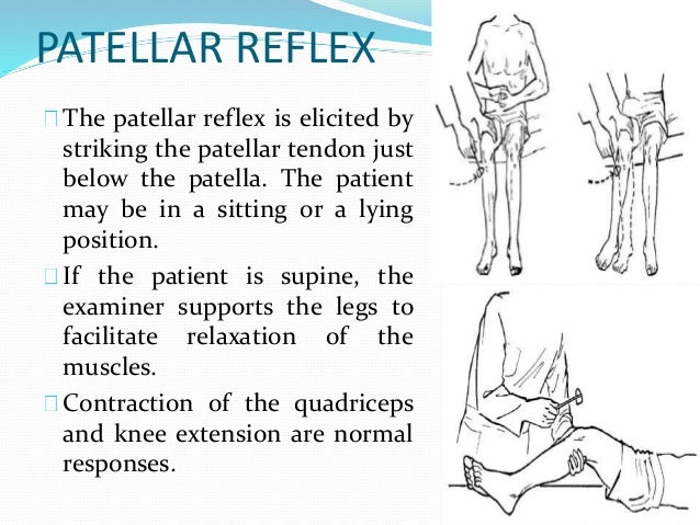
What muscles are involved in the patellar reflex?
The reflex arc of the Patellar reflex involves the femoral nerve and the spinal cord What is the effector muscle of the patellar reflex? the quadriceps femoris Calcaneal reflex Produced by gently striking the calcaneal tendon, results in plantar flexion of the foot The reflex arc of the calcaneal reflex involves the tibial nerve and the spinal cord
What are the effector muscles of the biceps reflex?
What are the effector muscles of the biceps reflex? The biceps brachii Triceps reflex Produced by striking the tendon of the triceps brachii near its insertion just proximal to the olecranon. Produces a slight twitch, or extension of the forearm at the elbow joint
What muscles are involved in the Achilles reflex?
and the Achilles reflex or ankle jerk reflex, mediated by S1 through the sciatic (tibial) nerve and elicited by tapping the tendon of the gastrocnemius muscle (Fig. 33.22D).
What is the calcaneal reflex response?
The Achilles reflex is a monosynaptic stretch reflex similar to the patellar reflex. In the Achilles reflex, the hammer taps the Achilles tendon while the foot is dorsiflexed, and the foot, in response, should jerk toward the plantar surface. The Achilles reflex originates in the S1 and S2 nerve roots.
What nerve is calcaneal reflex?
The LMN of the Achilles reflex consists of the ventral horn of the S1 nerve root and the tibial nerve.
Where is the calcaneal reflex?
0:110:59Achilles Heel Deep Tendon Reflex Test | Nursing Head to Toe ...YouTubeStart of suggested clipEnd of suggested clipWhich is located right above the heel.MoreWhich is located right above the heel.
How do you get your Achilles reflex?
How to Assess the Achilles Deep Tendon ReflexFind the achilles tendon.Locate the heel of the foot and dorsiflex the foot. The achilles is found right above the heel.Now have the patient dangle the foot while you dorsiflex it.While the foot is dorsiflex, tap with the reflex hammer briskly on the achilles tendon.
Where do you hit Achilles reflex?
Strike the tendon just above the calcaneus at the level of the malleoli. Standardize the size of reflex hammer used and force if needed. Palpate and visually observe the response to the provided stimulus (i.e., contraction of the calf and ankle plantar flexion). Repeat the procedure.
Which part of the reflex hammer will you use to test the calcaneal reflex?
Which part of the reflex hammer will you use to test the calcaneal reflex? The broad rubber side.
What is the receptor involved in the plantar reflex?
Plantar Reflex- The plantar reflex is elicited by stimulating the cutaneous receptors in the sole of the foot. In adults, stimulation of these receptors causes the toes to flex and move closer together.
What does absent Achilles reflex mean?
Your doctor will use a rubber hammer to tap firmly on the Achilles tendon, which connects the muscle at the back of your calf to your heel bone. In a normal test, your foot will move as though you were going to point your toes. A decreased or absent reflex may mean that there is compression in the S1 region.
What is the calcaneal reflex?
Calcaneal reflex. Produced by gently striking the calcaneal tendon, results in plantar flexion of the foot. The reflex arc of the calcaneal reflex involves. the tibial nerve and the spinal cord.
Which muscle produces plantar flexion?
the triceps brachii muscle. Plantar Reflex. Produced by applying pressure over the sole of the heal to the base of the large toe. Normal response is flexion (curling) of the toes and plantar flexion of the foot.
What is the Achilles reflex?
It is a type of stretch reflex that tests the function of the gastrocnemius muscle and the nerve that supplies it.
What is Grade 4 hyper reflexia?
Grade 4 ankle hyper reflexia is called ankle clonus. There is repetitive ankle dorsiflexion and plantarflexion on passive dorsiflexion of the foot by the examiner till the force applied by the examiner is withdrawn.
Can you dorsiflex your ankle?
Ankle of the patient is relaxed. It is helpful to support the ball of the foot at least somewhat to put some tension in the Achilles tendon, but don’t completely dorsiflex the ankle. A small strike is given on the Achilles tendon using a rubber hammer to elicit the response.
Is a deep tendon reflex a stretch reflex?
Being a deep tendon reflex, it is monosynaptic. It is also a stretch reflex. These are monosynaptic spinal segmental reflexes. When they are intact, integrity of the following is confirmed: cutaneous innervation, motor supply, and cortical input to the corresponding spinal segment.
Which muscles are involved in the plantar reflex?
effector muscles involved in the plantar reflex. gastrocnemius, soleus, and flexor digitorum longus. list the major events that occur in the patellar knee jerk reflex from the striking of the patellar ligament to the resulting response.
What is the function of the quadriceps femoris?
The quadriceps femoris is stretched, stimulating stretch receptors (muscle spindles) within the muscle. As a result, impulses pass along sensory neurons into the spinal cord and synapse with a motor neuron. Motor impulses travel out of the cord on nerve fibers that lead to the quadriceps femoris. Muscle fibers contract, and ...
What is the reflex arc?
A reflex arc refers to the neural pathway that a nerve impulse follows. The reflex arc typically consists of five components: A receptor, and independent sensory cell, or an ending of a sensory neuron, reacts to a stimulus (e.g., a stretch receptor). The sensory, or afferent, neuron sends a nerve impulse through an afferent pathway to ...
Where are polysynaptic reflexes found?
Polysynaptic reflexes are more complex, but also more common. They involve interneurons, found in the CNS , which further process stimulus and output information. Beyond simple reflexes with integrating centers in the spinal cord, more complex reflexes have integration centers in the brainstem or even in the cerebrum.
What muscle is a tap to the patellar tendon?
A tap to the patellar tendon stretches the quadriceps muscle (1) resulting in activation of the muscle spindle (2). The afferent neuron of the muscle spindle, detecting stretch, sends a signal to the spinal cord (3) and synapses directly with a motor neuron (4) that causes the quadriceps muscle to contract (5).
What is the purpose of the patellar reflex?
The primary purpose of the patellar reflex – the stretch reflex of the quadriceps femoris muscle – is to prevent excessive stretching of the quadriceps. The patellar reflex is illustrated in Figure 1.
Which neuron innervates contractile extrafusal fibers?
The sensory neuron synapses with a motor neuron in the spinal cord that innervates contractile extrafusal fibers. The contraction of the extrafusal fibers, that is, contraction of the belly of the muscle, releases tension on the intrafusal fibers, decreasing stimulation to neuron.
Which reflex is more complex, polysynaptic or monosynaptic?
This is the simplest reflex arc, and the integrating center is in the spinal cord. Polysyna ptic reflexes are more complex, but also more common.
What is reflex response?
A reflex is an involuntary (automatic) response to stimulus that quickly returns the body to homeostasis. There are several kinds of reflexes. Examples are shivering in response to low core body temperature; or withdrawing your hand from a hot stove when temperature and pain receptors in your hand register the stimulus.