What is the function of the corpora quadrigemina quizlet?
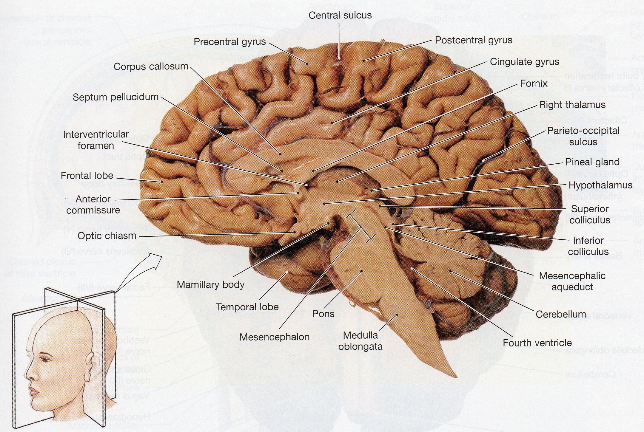
What are the functions of the corpora quadrigemina? SUPERIOR COLLICULI control reflexes of head & eyes in response to VISUAL stimuli. INFERIOR COLLICULI control reflexes of head & eyes in response to AUDITORY stimuli.
What is the function of the following structure corpora quadrigemina?
The corpora quadrigemina are four rounded elevations on the dorsal surface of the midbrain. The two superior colliculi are involved in visual reflexes and the two inferior colliculi are relay centres for auditory reflexes that operate when it is necessary to move the head to hear sounds more distinctly.
What is Corpus Quadrigemina?
The corpora quadrigemina (Latin for "quadruplet bodies", singular: corpus quadrigeminum) are the four colliculi, two inferior and two superior, that sit on the quadrigeminal plate on the posterior surface of the midbrain.
What part of the brain is corpora quadrigemina?
midbrainCorpora quadrigemina is the Latin terminology for the quadrigeminal bodies, also known as the colliculi. These round eminences are located on the posterior surface of the midbrain, just below the thalamus.
What structure of the corpora quadrigemina contains the auditory reflex center?
Answer and Explanation: The part of the brain stem that contains corpora quadrigemina, visual and auditory reflex center, is the midbrain.
Is corpora quadrigemina mammalian character?
The quadrigemina means quadruplet bodies. So, the small, solid and four lobes or colliculus called corpora quadrigemina are found in mammals. Thus, the correct answer is option A. i.e., Mammals.
What Is Corpora Quadrigemina?
Corpora quadrigemina are part of the midbrain. As the word ‘quad ’ stands for four, corpora quadrigemina is made up of four bodies. It is found in the midbrain's distal part. It is divided into two parts where the upper two calculi are called superior calculi and lower calculi are called inferior calculi.
Characteristics Of Corpora Quadrigemina
Corpora quadrigemina is the Latin term for the quadruple bodies, also known as the colliculi.
Superior Colliculi
The superior colliculi are made up of seven layers of grey and white matter that are arranged in seven levels from peripheral to deep:
Inferior Colliculi
The inferior colliculus has a grey matter nucleus in the centre. The nucleus has two zones i.e. dorsomedial and ventrolateral, that are surrounded by a dorsal cortex and an external cortex. The inferior colliculi receive auditory information from the cochlear nuclei via the terminal nerve fibres of the lateral lemniscus.
Things To Remember
Corporal Quadrigemina are the two pairs of colliculi on the dorsal surface of the midbrain composed of white matter externally and grey matter within
Which membrane separates the quadrigeminal cistern from the supracerebellar cister
An arachnoid membrane extending from the ascending part of the PPM to the vermis and, further, superiorly to the outer arachnoid membrane at the tentorial apex (i.e., the CPM) separates the quadrigeminal cistern from the supracerebellar cistern.
What is the posterior surface of the midbrain?
Posterior (Dorsal) Midbrain. The posterior surface of the adult midbrain is characterized by four elevations collectively called the corpora quadrigemina ( Fig. 13.4 ). The rostral two elevations are the superior colliculi, and the caudal two are the inferior colliculi. Just caudal to the inferior colliculus, the exit of ...
Which part of the brain contains the medulla, pons, and medulla?
It includes fibers descending to the spine (corticospinal), the medulla (corticobulbar), and the pons (corticopontine). The tegmentum of the midbrain contains all the ascending and many of the descending systems of the spinal cord or lower brainstem.
Where is the pons-midbrain junction?
Just caudal to the inferior colliculus, the exit of the trochlear nerve marks the pons-midbrain junction on the posterior surface of the brainstem, whereas the midbrain-diencephalic boundary is formed by the posterior commissure ( Fig. 13.5 ). Rostrolaterally, the inferior colliculus is joined to the medial geniculate body ...
Where are the peduncles located in the brain?
Degeneration of the substantia nigra is implicated in Parkinson's disease. Finally, the cerebral peduncles, located in the tegmentum, are bundles of nerves that run from the cerebrum to the spinal cord and are responsible for motor control. The pons is located between the midbrain and the medulla oblongata.
Which part of the brain is associated with vision?
The caudal pair is associated with acute hearing; the rostral pair is associated with vision. The most caudal portion of the brain is composed of the medulla oblongata as it tapers back into the spinal cord. On the ventral aspect of the brain are the origins of the cranial nerves Fig. 4-16 ).
What is the pineal gland?
On the midline, the pineal gland, a diencephalic structure, extends posteriorly above and between the superior colliculi ( Fig. 13.5 ). Tumors of the pineal may produce noncommunicating ...
