Autogenous bone grafts , also known as autografts, are made from your own bone, taken from somewhere else in the body. The bone is typically harvested from the chin, jaw, lower leg bone, hip, or the skull.
What are autogenous bone grafts?
Autogenous bone grafts or autografts are produced using your own bone, taken from other parts of the body. In this type of bone grafting, the surgeon may get bone from your jaw, chin, hip, lower leg bone, or skull. Autogenous bone grafts are beneficial since it uses a live bone, meaning it has living cell components that improve bone growth.
What is the difference between autograft and autologous graft?
allogeneic graftallograft. autodermic graft(autoepidermic graft) a skin grafttaken from the patient's own body. autologous graft(autoplastic graft) a graft taken from another area of the patient's own body; called also autograft. avascular grafta graft of tissue in which not even transient vascularization is achieved.
What is the difference between xenogenic and allogenic bone grafts?
Like an allogenic procedure, xenogenic grafts form a framework for bone from the encompassing region to promote bone growth and bone formation. Both xenogenic and allogenic bone grafting does not need a second procedure to gather your own bone, as with autografts.
What are the different types of bone grafts?
These include: Autogenous bone grafts or autografts are produced using your own bone, taken from other parts of the body. In this type of bone grafting, the surgeon may get bone from your jaw, chin, hip, lower leg bone, or skull.
What is a non-autogenous graft?
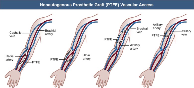
Note: The term “non-autogenous” is commonly understood to be any type of graft material that is not from the patient's body. In other words allogenic, alloplastic, allograft and xenograft materials are considered non-autogenous.
What is autogenous graft?
What is an Autogenous Bone Graft? Using the patient's own bone is called an autogenous bone graft. This means that at the time of surgery, the doctor makes an incision and takes a small piece of bone from an area of the mouth where it is not needed. In most cases, the bone is taken from a tooth extraction site.
What is Alloplast bone graft?
Alloplast. Alloplastic grafting material is synthetically derived or made from natural materials. The major advantages of alloplastic bone grafts include zero risk of disease transmission and low antigenicity. Alloplastic grafting materials include hydroxyapatite, dicalcium phosphates, and bioactive ceramics.
What does a dental bone graft do?
What is a dental bone graft? A dental bone graft adds volume and density to your jaw in areas where bone loss has occurred. The bone graft material may be taken from your own body (autogenous), or it may be purchased from a human tissue bank (allograft) or an animal tissue bank (xenograft).
Is autologous and autogenous the same?
Webster's Dictionary defines autogenous as “produced independently of external influence or aid” and autologous as “derived from the same individual involving one individual as both donor and recipient.”17 The Oxford English Dictionary defines autogenous as “self-produced, independent,” and autologous as “derived from ...
What is homograft skin graft?
Allograft, cadaver skin or homograft is human cadaver skin donated for medical use. Cadaver skin is used as a temporary covering for excised (cleaned) wound surfaces before autograft (permanent) placement. Cadaver skin is put over the excised wound and stapled in place.
What is the best dental bone graft material?
Hydroxyapatite is a synthetic bone graft, which is the most used now due to its osteoconduction, hardness, and acceptability by bone. Some synthetic bone grafts are made of calcium carbonate, which start to decrease in usage because it is completely resorbable in short time and makes breaking of the bone easier.
What is Alloplast made of?
Instead, the bone graft material alloplast is usually made from hydroxyapatite, which is a natural mineral that is the primary component of bone.
What are the different types of bone grafts?
Common options for bone grafting include:Xenograft Tissue.Alloplast Bone Graft.Autograft Tissue.Allograft Tissue.Growth Factors.
Is dental bone grafting painful?
Most patients who receive bone grafts are completely pain-free and do just fine as long as they take the antibiotics. Your dentist also has to wait for the bone graft to fuse with the natural bones that are already in your mouth.
Will gums grow over bone graft?
When a bone graft is needed for a dental implant, it is important that gum tissue does not grow over into the bone graft area. A piece of membrane material is placed over the area where the bone needs to be regenerated.
What is the success rate of bone grafts?
Composite bone grafts have 99.6% survival rate and 66.06% success rate. Allografts have 90.9% survival rate and 82.8% success rate.
What is an autodermic graft?
autodermic graft ( autoepidermic graft) a skin graft taken from the patient's own body.
What is a graft?
graft. [ graft] 1. any tissue or organ for implantation or transplantation. 2. to implant or transplant such tissues. This term is preferred over transplant in the case of skin grafts. See also implant. allogeneic graft allograft. autodermic graft ( autoepidermic graft) a skin graft taken from the patient's own body.
What is a homologous graft?
homologous graft a graft of tissue obtained from the body of another animal of the same species but with a genotype differing from that of the recipient; called also allograft and homograft.
What is a periosteal graft?
periosteal graft a piece of periosteum to cover a denuded bone.
What is a split skin graft?
split-skin graft ( split-thickness graft) a skin graft consisting of the epidermis and a portion of dermis. Diagram of a cross-section of the skin, demonstrating split thickness and full thickness skin grafts. From Roberts and Hedges, 1991.
What is an Ollier-Thiersch graft?
Ollier-Thiersch graft a very thin skin graft in which long, broad strips of skin, consisting of the epidermis, rete, and part of the corium, are used. omental graft a segment of omentum and its supplying vasculature, transplanted as a free flap to another area and revascularized by anastomosis of arteries and veins. pedicle graft pedicle flap.
What is a full thickness graft?
full-thickness graft a skin graft consisting of the full thickness of the skin, with little or none of the subcutaneous tissue.
What is autogenous graft?
Autogenous Grafts. Despite ongoing attempts to develop prosthetic or bioengineered materials for bypass grafting in the lower extremities, autologous vein graft remains the conduit of choice for infrainguinal revascularization. Though it was sporadically used for repair of popliteal aneurysms in the early 20th century, ...
What is the No Touch technique used in the harvest of autogenous vein?
Figure 92-2 “No-touch” technique used in the harvest of autogenous vein. To minimize spasm, papaverine (120 mg/L) is injected into the periadventitial plane before surgical exposure. Side branches are identified and ligated several millimeters away from the wall to prevent luminal narrowing after implantation and outward remodeling of the vein graft. Using a Silastic vessel loop to control the vein without direct manipulation with surgical instruments, the periadventitial tissues are divided and the vein is excised.
Which artery is used for distal anastomosis?
The posterior tibial, peroneal, and dorsalis pedis arteries provide the most straightforward options for placement of the distal anastomosis. Although the below-knee popliteal artery can also be used, fashioning the anastomosis can be problematic because of the nearly 90-degree angle that is formed between the graft and artery. With most hemodynamic analyses detailing an increase in flow separation and activation of proliferative metabolic pathways with right-angle anastomoses, grafts configured to this segment of the popliteal artery may benefit from excision and placement in an anatomic tunnel. Distal anastomoses to the above-knee popliteal artery do not offer the same constraints on their configuration; however, after mobilization of the proximal and distal graft, the segment that remains in situ can be quite short and of limited benefit.
What are the challenges of reversed configuration vein graft?
Challenges in constructing a reversed-configuration vein graft include significant proximal-to-distal tapering of a vein and the resulting size mismatch at the proximal or distal anastomoses (or both). Although use of an in situ grafting technique can be one solution to this issue, the combination of complete excision of the vein, lysis of the valves, and orientation in a nonreversed configuration offers an alternative technique. Despite the fact that initial attempts with this approach suggested results inferior to the in situ method, these early efforts were probably compromised by the use of an eversion valvulectomy technique and extensive injury to the vein wall. 100 With subsequent refinements in the approach to valve lysis, nonreversed vein grafts have proved as durable as other available techniques. 101–104
What is no touch technique?
92-2 ). 41–44 Though somewhat a misnomer, the “no-touch” technique involves limited handling of the vein without direct application of forceps or vascular clamps. Major branches are ligated away from the wall to avoid narrowing of the lumen and promote outward remodeling after implantation. Opinions regarding the importance of maintaining continuity of the vein during dissection versus early ligation for intermittent infusion of isotonic fluids are mixed. Despite not being studied in a directed manner, continued continuity of vein has been shown to reduce the formation of small thrombi on the wall, and cannulation and division of the distal vein promote the direct application of antithrombotic and smooth muscle relaxants directly into the lumen. Although limited vein graft handling is universally considered an important component of promoting both short- and long-term graft patency, this concept is challenged by the nearly identical clinical outcomes for reversed, nonreversed, and in situ vein grafting techniques. 45#N#,#N#46 If limiting vein manipulation is a dominant factor, one might intuitively expect improved outcomes with in situ grafting, where direct vein manipulation can be essentially eliminated, and compromised outcomes with nonreversed/excised grafts, which require complete mobilization with valvulotomy. The absence of such differences suggests that careful dissection of the vein is prudent, but absent extensive injury, it is of only modest importance to final outcomes.
What is the purpose of the great saphenous vein?
The concept of using the great saphenous vein as a graft with mobilization of only the proximal and distal segments while maintaining the interval region within its subcutaneous bed was initially suggested by Rob’s group in 1959. 105 Interested in the potential hemodynamic advantages offered by removal of the valves and retrograde perfusion of the conduit, Rob believed the procedure to be too time consuming for routine application. Hall, a visiting fellow from Norway, became intrigued by this approach, refined the techniques for routine clinical application, and published the first report of the in situ vein graft technique in 1962. 106 His version of the procedure, requiring valve excision by opening the vein at multiple locations along the graft, was tedious, required significant technical expertise, and failed to gain widespread acceptance. The development of efficient instrumentation for valve lysis provided the opportunity to reduce vein manipulation and maintain an intact vasa vasorum and fueled renewed enthusiasm for the in situ approach. Initial reports of this approach, championed by Leather et al, 107 suggested improved patency over excised vein grafts. 108 These early analyses are compromised by the use of historical controls, however, and contemporary comparisons of in situ versus reversed or nonreversed grafts fail to demonstrate significant differences in long-term outcomes. 46#N#,#N#103#N#,#N#109
Where do autogenous grafts come from?
Autogenous bone grafts or autografts are produced using your own bone, taken from other parts of the body. In this type of bone grafting, the surgeon may get bone from your jaw, chin, hip, lower leg bone, or skull. Autogenous bone grafts are beneficial since it uses a live bone, meaning it has living cell components that improve bone growth.
What are the drawbacks of autografts?
Nonetheless, one drawback to the autograft is that it involves a second method to reap bone from somewhere else in the body. Depending on your condition, a subsequent surgery may not be to your most significant advantage.
What is the procedure called when a cow is grafted?
In this type, very high temperatures are necessary to keep away from possible immune rejection and infection. Like an allogenic procedure, xenogenic grafts form a framework for bone from the encompassing region to promote bone growth and bone formation.
Why is bone grafting important?
Primary Importance of Bone Grafting. Bone grafting is a standard procedure to improve your smile and facial structure. They are a popular technique in the dental industry. However, their importance is not limited to dental use.
Why do you need a bone graft?
You may require bone grafting to advance bone healing and development for various medical reasons. Some particular conditions that may need a bone graft include: Diseases of the bone, such as osteonecrosis or cancer. An initial fracture that your doctor suspects will not recover without a graft.
What is the procedure to replace missing teeth?
Dental implant surgery, which you might need if you want to replace missing teeth. Surgically implanted appliances, as in all out-knee replacement, to help increase bone regeneration around the structure. With the use of the bone grafting technique, these conditions can be treated. Bone grafts provide a framework for the development of new, ...
What is the process of laying down new bone materials called?
Also called ossification , bone formation is laying down new bone materials by cells named osteoblast. Having new bone materials helps improve bone regeneration and growth.
As adjectives the difference between autologous and nonautologous
is that autologous is derived from part of the same individual (ie from the recipient rather than a different donor) while nonautologous is not autologous.
English
Derived from part of the same individual (i.e. from the recipient rather than a different donor).
