What does CSF consist of?
what does CSF consist of? Cerebrospinal fluid ( CSF) is a clear, colorless body fluid found in the brain and spinal cord. It is produced by specialised ependymal cells in the choroid plexuses of the ventricles of the brain, and absorbed in the arachnoid granulations. You may ask, Which diagnostic test evaluates cerebrospinal fluid?
Which structure forms the CSF?
Review of the literature Anatomy and physiology of cerebrospinal fluid
- Summary. ...
- Keywords. ...
- Comparative anatomy. ...
- The development of cerebrospinal fluid spaces retraces the steps of phylogenesis. ...
- Volumes. ...
- Cerebrospinal fluid secretion. ...
- Cerebrospinal fluid circulation. ...
- Cerebrospinal fluid absorption. ...
- The role of extra-arachnoid absorption pathways remains poorly elucidated. ...
What does CSF mean?
restoring confidence and relieving anxiety
- A. witless
- B. reassuring
- C. nasty
- D. hatched
What does CSF stand for in fluid?
What is the treatment for a cerebrospinal fluid (CSF) leak?
- Bed rest (days to up to 2 weeks)
- Hydration (2 to 3 quarts)
- IV caffeine infusions
- Saline infusions
What unit of measure is CSF?
Normal values typically range as follows: Pressure: 70 to 180 mm H2O. Appearance: clear, colorless. CSF total protein: 15 to 60 mg/100 mL.
What does CSF mean?
Cerebrospinal fluid (CSF) is a clear, colorless body fluid found within the tissue that surrounds the brain and spinal cord of all vertebrates. Cerebrospinal fluid. The cerebrospinal fluid circulates in the subarachnoid space around the brain and spinal cord, and in the ventricles of the brain.
How many mL is CSF?
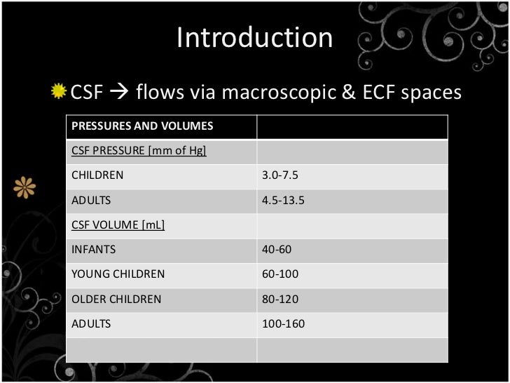
In normal adults, the CSF volume is 90 to 200 mL [1]; approximately 20 percent of the CSF is contained in the ventricles; the rest is contained in the subarachnoid space in the cranium and spinal cord. The normal rate of CSF production is approximately 20 mL per hour.
Why CSF test is done?
What is it used for? A CSF analysis may include tests to diagnose: Infectious diseases of the brain and spinal cord, including meningitis and encephalitis. CSF tests for infections look at white blood cells, bacteria, and other substances in the cerebrospinal fluid.
Where is the CSF located?
ventriclesCSF is secreted by the CPs located within the ventricles of the brain, with the two lateral ventricles being the primary producers. CSF flows throughout the ventricular system unidirectionally in a rostral to caudal manner.
Where is CSF present?
Summary. The cerebrospinal fluid (CSF) is contained in the brain ventricles and the cranial and spinal subarachnoid spaces. The mean CSF volume is 150 ml, with 25 ml in the ventricles and 125 ml in subarachnoid spaces. CSF is predominantly, but not exclusively, secreted by the choroid plexuses.
How much CSF is in a tube?
2-3 mlIdeally, CSF specimens should consist of these four (4) tubes each containing 2-3 ml of fluid.
How many drops of CSF is 1ml?
Please note: 1 mL of CSF is approximately equal to 25 drops from the Luer connector of the needle.
What is normal CSF pressure?
What is a Normal Opening Pressure? The normal range for CSF is reported differently in various sources, with most reporting a normal range of 7-18 cmH2O in adults,1 though some consider the normal range 5-25 cmH2O.
How much does a CSF test cost?
Cerebral Spinal Fluid (CSF) Test CostCityAverage PricePrice UptoMumbaiRs. 788.00Rs. 1500.00NalgondaRs. 440.00Rs. 600.00DelhiRs. 584.00Rs. 1500.00NoidaRs. 459.00Rs. 650.0010 more rows
What color is CSF fluid?
Color of the fluid—normal is clear and colorless. Changes in the color of the CSF are not diagnostic but may point to additional substances in the fluid. Yellow, orange, or pink CSF may indicate the breakdown of blood cells due to bleeding into the CSF or the presence of bilirubin.
What does CSF look like?
Cerebrospinal fluid (CSF) is a clear liquid that surrounds the brain and spinal cord.
What is the purpose of CSF testing?
CSF testing is performed to evaluate the level or concentration of different substances and cells in CSF in order to diagnose conditions affecting the brain and spinal cord (central nervous system).
Why is CSF testing important?
Any condition that disrupts this protective barrier may result in a change in the normal level or makeup of CSF. Because CSF surrounds the brain and spinal cord, testing a sample of CSF can be very valuable in diagnosing a variety of conditions affecting the central nervous system.
Why does my CSF pressure drop?
Decreased pressure may be due to dehydration, shock, or leakage of CSF through an opening (e.g., another lumbar puncture site or sinus fracture).
How much CSF is produced in the brain?
CSF is continually produced, circulated, and then absorbed into the blood. About 17 ounces (500 mL) of CSF are produced each day. This rate of production means that all the CSF is replaced every few hours.
When can you measure CSF pressure?
Pressure of the CSF can be measured when opening (starting) and closing (finishing) the collection.
When is CSF testing ordered?
CSF testing may be ordered when your healthcare practitioner suspects that you have a condition or disease involving your central nervous system. It may be ordered when: You have suffered trauma to the brain or spinal cord. You have been diagnosed with cancer that may have spread into the central nervous system.
What is the term for an infection of the brain and spinal cord?
Meningitis, an infection of the layers that cover the brain and spinal cord (meninges), and encephalitis, an infection in the brain. Autoimmune diseases that affect the central nervous system, such as multiple sclerosis. Cancers of the central nervous system or cancers that have spread to the central nervous system, such as leukemia.
What is a CSF cell count?
CSF cell count. Share. A CSF cell count is a test to measure the number of red and white blood cells that are in cerebrospinal fluid (CSF). CSF is a clear fluid that is in the space around the spinal cord and brain.
Why is CSF count important?
Why the Test is Performed. The CSF cell count may help detect: Meningitis and infection of the brain or spinal cord. Tumor, abscess, or area of tissue death (infarct) Inflammation.
What is the most common way to collect CSF?
A lumbar puncture (spinal tap) is the most common way to collect this sample. Rarely, other methods are used for collecting CSF such as:
Where is CSF found?
Cerebrospinal fluid ( CSF) is a clear, colorless body fluid found within the tissue that surrounds the brain and spinal cord of all vertebrates. It replaces the body fluid found outside the cells of all bilateral animals. The CSF is produced by specialised ependymal cells in the choroid plexuses of the ventricles of the brain, ...
What is the purpose of a CSF test?
Testing often includes observing the colour of the fluid, measuring CSF pressure , and counting and identifying white and red blood cells within the fluid; measuring protein and glucose levels; and culturing the fluid. The presence of red blood cells and xanthochromia may indicate subarachnoid hemorrhage; whereas central nervous system infections such as meningitis, may be indicated by elevated white blood cell levels. A CSF culture may yield the microorganism that has caused the infection, or PCR may be used to identify a viral cause. Investigations to the total type and nature of proteins reveal point to specific diseases, including multiple sclerosis, paraneoplastic syndromes, systemic lupus erythematosus, neurosarcoidosis, cerebral angiitis; and specific antibodies such as Aquaporin 4 may be tested for to assist in the diagnosis of autoimmune conditions. A lumbar puncture that drains CSF may also be used as part of treatment for some conditions, including idiopathic intracranial hypertension and normal pressure hydrocephalus.
How is CSF produced?
Firstly, a filtered form of plasma moves from fenestrated capillaries in the choroid plexus into an interstitial space , with movement guided by a difference in pressure between the blood in the capillaries and the interstitial fluid. This fluid then needs to pass through the epithelium cells lining the choroid plexus into the ventricles, an active process requiring the transport of sodium, potassium and chloride that draws water into CSF by creating osmotic pressure. Unlike blood passing from the capillaries into the choroid plexus, the epithelial cells lining the choroid plexus contain tight junctions between cells, which act to prevent most substances flowing freely into CSF. Cilia on the apical surfaces of the ependymal cells beat to help transport the CSF.
What causes CSF to leak?
CSF can leak from the dura as a result of different causes such as physical trauma or a lumbar puncture, or from no known cause when it is termed a spontaneous cerebrospinal fluid leak. It is usually associated with intracranial hypotension: low CSF pressure. It can cause headaches, made worse by standing, moving and coughing, as the low CSF pressure causes the brain to "sag" downwards and put pressure on its lower structures. If a leak is identified, a beta-2 transferrin test of the leaking fluid, when positive, is highly specific and sensitive for the detection for CSF leakage. Medical imaging such as CT scans and MRI scans can be used to investigate for a presumed CSF leak when no obvious leak is found but low CSF pressure is identified. Caffeine, given either orally or intravenously, often offers symptomatic relief. Treatment of an identified leak may include injection of a person's blood into the epidural space (an epidural blood patch ), spinal surgery, or fibrin glue.
How does CSF protect the brain?
Protection: CSF protects the brain tissue from injury when jolted or hit, by providing a fluid buffer that acts as a shock absorber from some forms of mechanical injury. Prevention of brain ischemia: The prevention of brain ischemia is aided by decreasing the amount of CSF in the limited space inside the skull.
How much cerebrospinal fluid is produced in the brain?
The brain produces roughly 500 mL of cerebrospinal fluid per day , at a rate of about 25 mL an hour. This transcellular fluid is constantly reabsorbed, so that only 125–150 mL is present at any one time. CSF volume is higher on a mL/kg basis in children compared to adults.
What test is used to detect a CSF leak?
If a leak is identified, a beta-2 transferrin test of the leaking fluid, when positive, is highly specific and sensitive for the detection for CSF leakage. Medical imaging such as CT scans and MRI scans can be used to investigate for a presumed CSF leak when no obvious leak is found but low CSF pressure is identified.
How much CSF is produced in a day?
CSF is produced at a rate of 0.2–0.7 mL per minute or 500–700 mL per day.1The main function of the CSF is to reduce buoyancy of the brain. It also supplies nutrients as well as helps in removal of various substances like amino acids, neurotransmitters, metabolic byproducts and cells.
Why is CSF analysis important?
Hence analysis of CSF by various methods will help in diagnosis as well as prognostication and response to therapy . CSF analysis is particularly useful in various acute neurological conditions and helps in rapid diagnosis of the conditions and initiate therapeutic measures .
What is cerebral fluid?
Cerebrospinal fluid (CSF) is a clear fluid circulating in the intracranial and spinal compartments. Under normal conditions, the composition of CSF remains constant. However, in various neurological disease especially in acute conditions, the composition, quantity and its pressure can be altered. By measuring the levels ...
What are the indications for LP and CSF?
Patients with suspected meningitis is one of the major indication for LP and CSF study. Meningitis can be community acquired or hospital acquired and caused by various micro organisms ranging from bacteria, virus, fungus, protozoa, etc.9,10Aseptic meningitis is a condition that needs to be distinguished from other forms of meningitis that need a CSF analysis.11Presentation of meningitis varies from acute debilitating illness or chronic symptoms as in tuberculosis. Patients suspected to have acute meningitis usually present with altered consciousness, fever and neck stiffness. The classic triad is seen only in 46% of patients. In others one or two of the signs of triad may be present. In addition patients can present with nausea, vomiting, headache and photophobia. In patients with meningoencephalitis additional clinical signs at presentation include altered sensorium, confusion, behavioural changes, seizures, focal neurological deficits.
Where is cerebral fluid secreted?
CSF is present in both the intracranial and spinal compartments. It is continuously being secreted by the choroid plexus at a constant rate inside the ventricles of the brain and circulates in the subarachnoid space of the brain ...
Can a manometer be used to measure CSF?
A manometer can be connected if CSF opening pressure measurement is planned. Color of CSF is noted and if blood stained due to traumatic puncture, it may be needed to wait for the blood to be cleared before samples are collected. Samples are usually collected in three to four test tubes each of 3–5 mL CSF for analysis.
Viral (aseptic) meningitis
WBC: elevated (50 – 1000 cells/µL, primarily lymphocytes, can be PMN early on)
Subarachnoid haemorrhage
Appearance: blood-stained initially, then xanthochromia (yellowish) >12 hours later
Case 1
A 55-year-old woman has become increasingly more confused over the last 2 months. Over the last 3 days, she has been vomiting and suffering from lack of energy. She has no neck stiffness and a CD4 count of 100/mm³
Case 2
A 28-year-old male presents with a 12-hour history of high fever, severe headache, confusion, photophobia and neck stiffness. He has no significant past medical history and takes no regular medication.
Case 3
A 38-year-old female presents with 24 hours of headache, photophobia and mild neck stiffness, in addition to coryzal symptoms. She is fully orientated and her observations are stable.
Case 4
A 52-year-old male presents to A&E with history of a sudden onset severe headache which occurred whilst he was at his desk yesterday. Since the headache, he has been feeling nauseated, but he is otherwise well and fully orientated. Examination is largely unremarkable, but he does appear to have some mild neck stiffness.
Latest Podcast
In this episode, we chat to Dr Dan Magnus, a Paediatric Emergency Consultant, about his work in a children's A&E department and his interests in global health. Dr Magnus takes us through the different training pathways, the best and hardest bits of the job, and gives some inspiring advice for anyone who feels they haven't got it all sorted yet.
Why are CSFdisbursements important?
Historically, the CSFdisbursements remained crucial for building foreign currency reserves, lessening strains on the external accounts and narrowing down the budget deficit.
When was Davao del Sur's credit surety fund launched?
Davao del Sur's credit surety fund is first to be launched in 2018
Overview
Cerebrospinal fluid (CSF) is a clear, colorless body fluid found within the tissue that surrounds the brain and spinal cord of all vertebrates.
CSF is produced by specialised ependymal cells in the choroid plexus of the ventricles of the brain, and absorbed in the arachnoid granulations. There is about 125 mL of CSF at any one time, and about 500 mL is generated every day. CSF acts as a cushion or buffer, providing basic mechanic…
Structure
There is about 125–150 mL of CSF at any one time. This CSF circulates within the ventricular system of the brain. The ventricles are a series of cavities filled with CSF. The majority of CSF is produced from within the two lateral ventricles. From here, CSF passes through the interventricular foramina to the third ventricle, then the cerebral aqueduct to the fourth ventricle. From the fourth ventricle, the fluid passes into the subarachnoid space through four openings – the central canal o…
Development
At around the third week of development, the embryo is a three-layered disc, covered with ectoderm, mesoderm and endoderm. A tube-like formation develops in the midline, called the notochord. The notochord releases extracellular molecules that affect the transformation of the overlying ectoderm into nervous tissue. The neural tube, forming from the ectoderm, contains CSF prior to the development of the choroid plexuses. The open neuropores of the neural tube close after the …
Physiology
CSF serves several purposes:
1. Buoyancy: The actual mass of the human brain is about 1400–1500 grams; however, the net weight of the brain suspended in CSF is equivalent to a mass of 25-50 grams. The brain therefore exists in neutral buoyancy, which allows the brain to maintain its density without being impaired by its own weight, which would cut off blood supply and kill neurons in the lower sections without C…
Clinical significance
CSF pressure, as measured by lumbar puncture, is 10–18 cmH2O (8–15 mmHg or 1.1–2 kPa) with the patient lying on the side and 20–30 cmH2O (16–24 mmHg or 2.1–3.2 kPa) with the patient sitting up. In newborns, CSF pressure ranges from 8 to 10 cmH2O (4.4–7.3 mmHg or 0.78–0.98 kPa). Most variations are due to coughing or internal compression of jugular veins in the neck. When lying down, the CSF pressure as estimated by lumbar puncture is similar to the intracrania…
History
Various comments by ancient physicians have been read as referring to CSF. Hippocrates discussed "water" surrounding the brain when describing congenital hydrocephalus, and Galen referred to "excremental liquid" in the ventricles of the brain, which he believed was purged into the nose. But for some 16 intervening centuries of ongoing anatomical study, CSF remained unmentioned in the literature. This is perhaps because of the prevailing autopsy technique, whic…
Other animals
During phylogenesis, CSF is present within the neuraxis before it circulates. The CSF of Teleostei fish is contained within the ventricles of the brains, but not in a nonexistent subarachnoid space. In mammals, where a subarachnoid space is present, CSF is present in it. Absorption of CSF is seen in amniotes and more complex species, and as species become progressively more complex, the system of absorption becomes progressively more enhanced, and the role of spinal epidural …
See also
• Neuroglobin
• Pandy's test
• Reissner's fiber
• Syrinx (medicine)