How is the third ventricle connected to the lateral ventricles?
The third ventricle is connected to the lateral ventricles by channels called interventricular foramina or foramina of Monro. These channels allow cerebrospinal fluid to flow from the lateral ventricles to the third ventricle. The cerebral aqueduct connects the third ventricle to the fourth ventricle.
What is an enlarged third ventricle associated with?
An enlarged third ventricle has been associated with psychiatric illnesses such as schizophrenia; however, the accuracy of this theory hasn't been conclusively proven. Medically reviewed by Healthline's Medical Network on March 9, 2015.
What is the floor of the third ventricle made of?
The floor of the third ventricle is formed by a number of structures including the hypothalamus, subthalamus, mammilary bodies, infundibulum (pituitary stalk), and the tectum of the midbrain. The lateral walls of the third ventricle are formed by the walls of the left and right thalamus.
See more
What surrounds the 3rd ventricle?
The lateral walls of the third ventricle are formed by the walls of the left and right thalamus. The anterior wall is formed by the anterior commissure (white matter nerve fibers), lamina terminalis, and optic chiasma. The posterior wall is formed by the pineal gland and habenular commissures.
Does the thalamus encloses the third ventricle?
Location of the thalamus The thalamus surrounds the third ventricle. It is a subdivision of part of the brain called the diencephalon and is one of the largest structures derived from the diencephalon during embryonic development.
What forms the walls of the 3rd ventricle?
Much of the medial surface of the thalamus and hypothalamus form the wall of the third ventricle. Part of the hypothalamus forms its floor. Its thin, membranous roof contains the choroid plexus. The third ventricle narrows quickly at the posterior end of the mammillary bodies.
What structure borders on the third ventricle?
The third ventricle is bordered anteriorly by the lamina terminalis. Its inferior border is the ventral diencephalon (VDC). Its lateral border is made up of the hypothalamus and other VDC structures (ventrally) and the thalamus (dorsally).
What is thalamus and hypothalamus?
The thalamus is a small structure, located right above the brainstem responsible for relaying sensory information from the sense organs. The hypothalamus is a small and essential part of the brain, located precisely below the thalamus. It is responsible for transmitting motor information for movement and coordination.
What surrounds the thalamus?
Furthermore, the thalami are each surrounded two layers of white matter. Dorsally, it is covered by a layer known as the stratum zonale; while laterally, it is covered by the external medullary lamina, which separates the lateral and ventral thalamus from the thalamic reticular nucleus and the subthalamus.
What is the 3rd ventricle of the brain called?
StructureNameFromTocerebral aqueduct (Sylvius)third ventriclefourth ventriclemedian aperture (Magendie)fourth ventriclesubarachnoid space via the cisterna magnaright and left lateral aperture (Luschka)fourth ventriclesubarachnoid space via the cistern of great cerebral vein1 more row
What is the name of the structure that connects the third and fourth ventricle?
The third ventricle is connected to the fourth ventricle via the cerebral aqueduct (also called the aqueduct of Sylvius).
What is foramen of Magendie?
Medical Definition of foramen of Magendie : a passage through the midline of the roof of the fourth ventricle of the brain that gives passage to the cerebrospinal fluid from the ventricles to the subarachnoid space.
What is the third ventricle?
The third ventricle is one of the four ventricles in the brain that communicate with one another. As with the other ventricles of the brain, it is filled with cerebrospinal fluid, which helps to protect the brain from injury ...
Where is the third ventricle located?
The third ventricle is a narrow cavity that is located between the two halves of the brain. The third ventricle sends messages to and receives messages from the lateral ventricles, which are located in front of the third ventricle, and the aqueduct of the midbrain, which is located directly behind the third ventricle.
What are the abnormalities of the third ventricle?
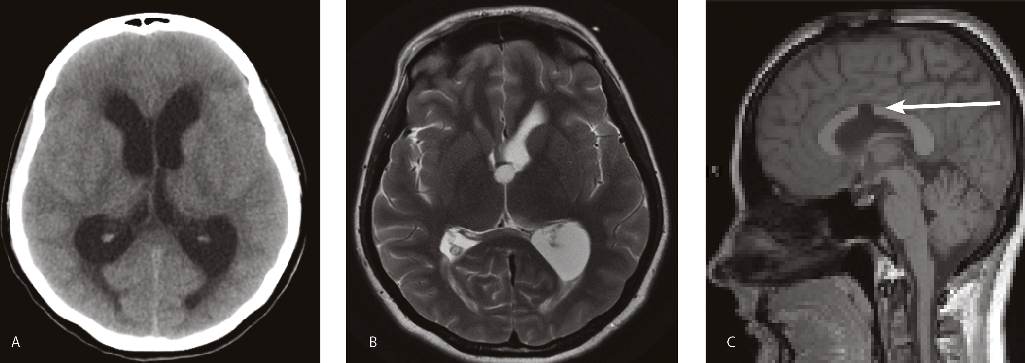
Abnormalities of the third ventricle are associated with various conditions including hydrocephalus, meningitis, and ventriculitis. Hydrocephalus is an excessive buildup of fluid on the brain. Meningitis is inflammation of the membranes that cover the brain and spinal cord, whereas ventriculitis is an inflammatory condition of the ventricles.
Can meningitis cause ventriculitis?
Meningitis and ventriculitis can both be caused by trauma to a ventricle, including the third ventricle, althought traumatic meningitis is rare. An enlarged third ventricle has been associated with psychiatric illnesses such as schizophrenia; however, the accuracy of this theory hasn’t been conclusively proven.
Where is the third ventricle located?
The third ventricle is situated between the right and the left thalamus. It has two protrusions on its top surface—the supra-optic recess (located above the optic chiasm) and the infundibular recess (located above the optic stalk).
What is the function of the third ventricle?
Similar to the other brain ventricles, the main function of the third ventricle is to produce, secrete, and convey CSF. It also has several very important secondary roles, such as protection of the brain from trauma and injury and transport of nutrients and waste from the body’s central nervous system. 1
How many walls does the third ventricle have?
The third ventricle is a cuboid-shaped structure that has a roof, floor, and four walls—the anterior, posterior, and two lateral walls, respectively.
What ventricle is affected by stroke?
Stroke: The third ventricle can be affected by the bleeding in the brain that occurs when a person has a stroke.
What causes the third ventricle to be misshapen?
Congenital malformations: Hereditary conditions can cause the third ventricles to become misshapen.
Which ventricle is the main site for CSF production?
The third ventricle is the main site for CSF production. CSF has three main roles in the brain:
What causes enlargement of the third ventricle?
Congeni tal conditions: Genetic malformations such as congenital aqueductal stenosis can cause enlargement of the third ventricle. 5
What is the third ventricle?
Key Takeaways. The third ventricle is one of four brain ventricles. It is a cavity filled with cerebrospinal fluid located between the two hemispheres of the diencephalon of the forebrain. The third ventricle helps to protect the brain from trauma and injury. The third ventricle is also involved in the transport of both nutrients and waste from ...
Which ventricle is connected to the third ventricle?
These channels allow cerebrospinal fluid to flow from the lateral ventricles to the third ventricle. The cerebral aqueduct connects the third ventricle to the fourth ventricle. The third ventricle also has small indentations known as recesses.
How many components are there in the third ventricle?
The third ventricle can be described as having six components: a roof, a floor, and four walls. The roof of the third ventricle is formed by a part of the choroid plexus known as the tela chorioidea. The tela chorioidea is a dense network of capillaries that is surrounded by ependymal cells. These cells produce cerebrospinal fluid.
What are the structures that make up the third ventricle?
The floor of the third ventricle is formed by a number of structures including the hypothalamus, subthalamus, mammilary bodies, infundibulum (pituitary stalk), and the tectum of the midbrain. The lateral walls of the third ventricle are formed by the walls of the left and right thalamus.
What is the function of the diencephalon?
The diencephalon is a division of the forebrain that relays sensory information between brain regions and controls many autonomic functions. It links endocrine system, nervous system, and limbic system structures. The third ventricle can be described as having six components: a roof, a floor, and four walls. The roof of the third ventricle is ...
What causes a dilated third ventricle?
Third ventricle issues and abnormalities can occur in a variety of conditions like stroke, meningitis and hydrocephalus. A relatively common cause of an abnormality of the third ventricle occurs with congenital hydrocephalus (abnormal contour with a dilated third ventricle).
Where is the third ventricle located?
Updated July 05, 2019. The third ventricle is a narrow cavity located between the two hemispheres of the diencephalon of the forebrain. The third ventricle is part of a network of linked cavities (cerebral ventricles) in the brain that extend to form the central canal of the spinal cord.
What is the floor of the third ventricle?
The floor of the third ventricle is formed by hypothalamic structures and this can be opened surgically between the mamillary bodies and the pituitary gland in a procedure called an endoscopic third ventriculostomy. An endoscopic third ventriculostomy can be performed in order to release extra fluid caused by hydrocephalus .
Where does the third ventricle originate?
The third ventricle, like other parts of the ventricular system of the brain, develops from the neural canal of the neural tube. Specifically, it originates from the most rostral portion of the neural tube which initially expands to become the prosencephalon. The lamina terminalis is the rostral termination of the neural tube.
What is the lateral side of the ventricle?
The lateral side of the ventricle is marked by a sulcus – the hypothalamic sulcus – from the inferior side of the interventricular foramina to the anterior side of the cerebral aqueduct. The lateral border posterior/superior of the sulcus constitutes the thalamus, while anterior/inferior of the sulcus it constitutes the hypothalamus. The interthalamic adhesion usually tunnels through the thalamic portion of the ventricle, joining together the left and right halves of the thalamus, although it is sometimes absent, or split into more than one tunnel through the ventricle; it is currently unknown whether any nerve fibres pass between the left and right thalamus via the adhesion (it has more resemblance to a herniation than a commissure ).
What is the slit in the diencephalon?
It is a slit-like cavity formed in the diencephalon between the two thalami, in the midline between the right and left lateral ventricles, and is filled with cerebrospinal fluid (CSF). Running through the third ventricle is the interthalamic adhesion, which contains thalamic neurons and fibers that may connect the two thalami.
Which part of the ventricle is responsible for sleep?
The superior part of the posterior border constitutes the habenular commissure, while more centrally it the pineal gland, which regulates sleep and reacts to light levels.
Which bone distends towards the parietal bone?
Caudal of the bend, the ventricle border forms the epithalamus, and begins to distend towards the parietal bone (in lower vertebrates, it distends more specifically to the parietal eye ); the border of the distention forms the pineal gland.
What is a rare tumor that can arise in the third ventricle?
A chordoid glioma is a rare tumour that can arise in the third ventricle.

Anatomy
- Structure
The third ventricle is a cuboid-shaped structure that has a roof, floor, and four walls—the anterior, posterior, and two lateral walls, respectively. The roof is made up of the choroid plexus where CSF is produced by ependymal cells. The floor is made up of the hypothalamus, subthalamus, mam… - Location
The third ventricle is a midline structure. It is found between the cerebral hemispheres. It communicates directly with each lateral ventricle via the foramen of Monro and with the fourth ventricle via the aqueduct of Sylvius.2 The third ventricle is situated between the right and the lef…
Anatomical Variations
- There are several variations of the third ventricle.3The most common variations are: 1. Masses: Deformities of the different segments of the floor can be caused by tumors of the posterior fossa and hydrocephalus. 2. Longstanding hydrocephalus and increased intracranial pressure: The third ventricle is a common site for anatomical variations in people with congenital hydrocephalus, a …
Function
- The third ventricle is the main site for CSF production. CSF has three main roles in the brain: 1. Protection: CSF acts as a cushion for the brain, limiting neural damage in cranial injuries. 2. Buoyancy:CSF allows structures to float in the brain. By being immersed in CSF, the net weight of the brain is reduced to approximately 25 grams, preventing excessive pressure on the brain. 3. C…
Associated Conditions
- Abnormalities of the third ventricle are associated with other medical conditions. Some of the most common conditions associated with the third ventricle are: 1. Hydrocephalus: Hydrocephalus is a condition that leads to excessive buildup of CSF in and around the brain. In children, it can cause progressive enlargement of the head, potentially causing convulsions, tunn…
Tests
- Ventriculomegaly can be detected through prenatal tests or after the baby is born. Tests include: 1. Prenatal ultrasound 2. Amniocentesis 3. Magnetic resonance imaging (MRI) In adults, if there is suspicion of a tumor, hydrocephalus, or congenital malformation, a doctor may use the following to help diagnose the condition: 1. Physical examination 2. Eye examination 3. CT scan 4. MRI sc…
Overview
The third ventricle is one of the four connected ventricles of the ventricular system within the mammalian brain. It is a slit-like cavity formed in the diencephalon between the two thalami, in the midline between the right and left lateral ventricles, and is filled with cerebrospinal fluid (CSF).
Running through the third ventricle is the interthalamic adhesion, which contains thalamic neurons and fibers that may connect the two thalami.
Structure
The third ventricle is a narrow, laterally flattened, vaguely rectangular region, filled with cerebrospinal fluid, and lined by ependyma. It is connected at the superior anterior corner to the lateral ventricles, by the interventricular foramina, and becomes the cerebral aqueduct (aqueduct of Sylvius) at the posterior caudal corner. Since the interventricular foramina are on the lateral edge, the corner o…
Development
The third ventricle, like other parts of the ventricular system of the brain, develops from the neural canal of the neural tube. Specifically, it originates from the most rostral portion of the neural tube which initially expands to become the prosencephalon. The lamina terminalis is the rostral termination of the neural tube. After about five weeks, different portions of the prosencephalon begin to take distinct developmental paths from one another – the more rostral portion become…
Clinical significance
The floor of the third ventricle is formed by hypothalamic structures and this can be opened surgically between the mamillary bodies and the pituitary gland in a procedure called an endoscopic third ventriculostomy. An endoscopic third ventriculostomy can be performed in order to release extra fluid caused by hydrocephalus.
Several studies have found evidence of ventricular enlargement to be associated with major depr…
See also
• Biology of depression
• Suprapineal recess
• Tanycytes line the bottom of the ventricle
External links
• Stained brain slice images which include the "third%20ventricle" at the BrainMaps project