What does hyperechoic and hypoechoic mean?
Ultrasound terms:
- Hyperechoic – more echogenic (brighter) than normal
- Hypoechoic – less echogenic (darker) than normal
- Isoechoic – the same echogenicity as another tissue
What causes hypodense lesions in the liver?
What Are Liver Lesions?
- Symptoms. Benign liver lesions usually don’t cause any symptoms. ...
- Diagnosis. Blood tests: They might use these to test for viral hepatitis or to see how well your liver is working.
- Treatment. If you don’t have any symptoms, you may not need to do anything about the lesion. ...
What is increased echogenicity mean?
What does increased echogenicity mean? Increased echogenicity means that on a sonogram study the liver had a lighter or whiter appearance than is typical. This is called fatty infiltration and can be caused by many things such as medications like cholesterol lowering drugs and many other medications.
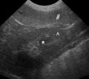
What is a hypoechoic lesion in the liver?
Hyperechoic liver lesions. A hyperechoic liver lesion on ultrasound can arise from a number of entities, both benign and malignant. A benign hepatic hemangioma is the most common entity encountered, but in patients with atypical findings or risk for malignancy, other entities must be considered.
What does hyperechoic mean on liver ultrasound?
A hyperechoic liver lesion on ultrasound can arise from a number of entities, both benign and malignant. A benign hepatic hemangioma is the most common entity encountered, but in patients with atypical findings or risk for malignancy, other entities must be considered. Benign.
What does hypoechoic liver mean?
A hypoechoic mass may be a tumor or abnormal growth. It may be benign or malignant. A benign tumor may grow but it will not spread (metastasize) to other organs. A malignant (cancerous) tumor can spread and invade other parts of the body.
Should I be worried about liver lesions?
Liver lesions are abnormal growths that may be noncancerous (benign) or cancerous. Benign lesions occur for a variety of reasons and are typically not cause for concern. Liver cancer is less common but more serious.
Is fatty liver hypoechoic or hyperechoic?
On ultrasound, fatty liver is hyperechoic compared with renal cortex and spleen, whereas fatty sparing is isoechoic or hypoechoic. On unenhanced computed tomography (CT), liver density less than 40 Hounsfield units (HU) [5] or a density difference of more than 10 HU between spleen and liver indicates fatty liver [6].
What does hyperechoic indicate?
Hyperechoic. This term means "lots of echoes." These areas bounce back many sound waves. They appear as light gray on the ultrasound. Hyperechoic masses are not as dense as hypoechoic ones are. They may contain air, fat, or fluid.
Is hypoechoic or hyperechoic better?
Hyperechoic tissues generate a greater echo usually displaying as lighter colors during ultrasound imaging. Hypoechoic – Refers to structures that create weaker echoes such as a fluid. Tissues with lower echogenicity are usually represented as darker colors on ultrasound.
How do you know if a liver lesion is cancerous?
Malignant liver lesions are diagnosed in a myriad of ways. If your healthcare provider suspects you have liver cancer, any of these may be ordered: Blood tests like alpha-fetoprotein (AFP) tumor marker and liver function tests (LFTs) Imaging tests like ultrasounds, computerized tomography (CT) scans, and MRIs.
Can a CT scan tell if a tumor is benign?
Cysts that appear uniform after examination by ultrasound or a computerized tomography (CT) scan are almost always benign and should simply be observed. If the cyst has solid components, it may be benign or malignant and should have further evaluation.
What is the difference between a lesion and a tumor on the liver?
Liver lesions are groups of abnormal cells in your liver. Your doctor may call them a mass or a tumor. Noncancerous, or benign, liver lesions are common. They don't spread to other areas of your body and don't usually cause any health issues.
How long does it take to reverse a fatty liver?
If you have fatty liver disease, the damage may be reversed if you abstain from alcohol for at least 2 weeks. After this point, it's usually safe to start drinking again if you stick to the NHS guidelines on alcohol consumption.
Can fatty liver Be Reversed?
It can lead to much more serious conditions including cirrhosis and liver failure.” The good news is that fatty liver disease can be reversed—and even cured—if patients take action, including a 10% sustained loss in body weight.
Can a hypoechoic mass be benign?
The echo texture of a benign mass will usually be homogeneous with an isoechoic, hyperechoic, to mildly hypoechoic echogenicity. Some benign lesions will also exhibit mild acoustic enhancement on ultrasound, and might be slightly compressible.
What is hyperechoic on ultrasound?
2 Answers. “Hyperechoic” is a word used to describe a particularly dark area that shows up on an ultrasound. Dense tissue and bone will usually show up as being hyperechoic.
What causes fatty liver?
The condition can be caused by any of the following: Excessive alcohol consumption ( this is the most common cause) Diabetes. Obesity. Hypertension (high blood pressure) HIV. Inflammatory bowel disease. Poor nutrition.
What is a benign liver lesion on ultrasound?
A hyperechoic liver lesion on ultrasound can arise from a number of entities, both benign and malignant. A benign hepatic hemangioma is the most common entity encountered, but in patients with atypical findings or risk for malignancy, other entities must be considered.
Is cholangiocarcinoma echogenic?
The presence of hyperechogenicity can be a result of fat within a liver lesion 2, although some non-fat-containing lesions may also be echogenic (e.g. hepati c hemangioma). On this page: Article: Radiographic features.