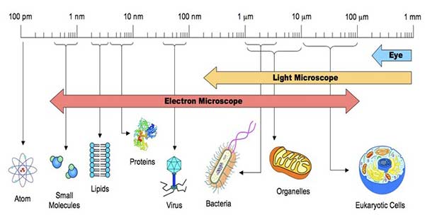
How do you measure the size of a paramecium? Then, divide 1,400 microns by this number to obtain an estimate of the cell's size in microns. For example, suppose it takes 8 paramecia laid end to end to equal the diameter of the field of view. If you divide 1,400 by 8, you get 175. Thus, the size of a single paramecium is approximately 175 microns.
See more
How do you determine the size of an organism under a microscope?
Divide the number of cells in view with the diameter of the field of view to figure the estimated length of the cell. If the number of cells is 50 and the diameter you are observing is 5 millimeters in length, then one cell is 0.1 millimeter long. Measured in microns, the cell would be 1,000 microns in length.
How do you find the actual size of a specimen?
To calculate the actual size of a magnified specimen, the equation is simply rearranged: Actual Size = Image size (with ruler) ÷ Magnification.
What is the size of a Paramecium in micrometers?
Species of Paramecium range in size from 50 to 330 micrometres (0.0020 to 0.0130 in) in length. Cells are typically ovoid, elongate, foot- or cigar-shaped.
How do you calculate cell size?
Divide the number of cells that cross the diameter of the field of view into the diameter of the field of view to figure out the length of one cell. If the diameter of the field is 5mm and you estimate that 50 cells laid end to end will cross the diameter, then 5mm/50 cells is 0.1mm/cell.
How do you determine the size of an object?
8:148:32Calculating Size of an Object - YouTubeYouTubeStart of suggested clipEnd of suggested clipSo that's just a little something extra but this is how you do it this is how you calculate the sizeMoreSo that's just a little something extra but this is how you do it this is how you calculate the size of an object.
How do you estimate the size of a microscopic specimen?
Estimating the Size of the Specimen Under Observation Remember that 1 μm = 0.001 mm. To estimate the size of an object seen with a microscope, first estimate what fraction of the diameter of the field of vision that the object occupies. Then multiply the diameter you calculated in micrometers by that fraction.
What is the largest paramecium?
multimicronucleatum is the largest species and is slimmer and more pointed than P. caudatum. It has one macronucleus and 3 or 4 micronuclei.
How do you identify a paramecium?
Surprisingly, paramecium is visible to the naked eye and has an elongated slipper like shape, that's the reason it's also referred to as a slipper animalcule. The posterior end of the body is pointed, thick and cone-like while the anterior part is broad and blunt. The widest part of the body is below the middle.
What is fit number microscope?
The field number (FN) in microscopy is defined as the diameter of the area in the intermediate image plane that can be observed through the eyepiece. A field number of, e.g., 20 mm indicates that the observed sample area after magnification by the objective lens is restricted to a diameter of 20 mm.
How do you find the length of a cell in micrometers?
1:443:49Calculating cell size when looking through a microscope - YouTubeYouTubeStart of suggested clipEnd of suggested clipHere so to get to micrometers. I just move the decimal place one two three places and that'll giveMoreHere so to get to micrometers. I just move the decimal place one two three places and that'll give me 90. So 90 my 90 micrometers would be about the length of one onion so in this example.
How do you determine the size of a cell with a scale bar?
Scale barMeasure the scale bar image (beside drawing) in mm.Convert to µm (multiply by 1000).Magnification = scale bar image divided by actual scale bar length (written on the scale bar).
How do you find the actual size of a magnified cell?
Calculating Magnification & Specimen Size: BasicsMagnification = image size / actual size.Actual size = image size / magnification.Image size = magnification x actual size.
How big is a paramecium?
Description. Species of Paramecium range in size from 50 to 330 micrometres (0.0020 to 0.0130 in) in length. Cells are typically ovoid, elongate, foot- or cigar-shaped. The body of the cell is enclosed by a stiff but elastic structure called the pellicle. This consists of the outer cell membrane (plasma membrane), ...
What is the function of the paramecium?
The macronucleus controls non-reproductive cell functions, expressing the genes needed for daily functioning. The micronucleus is the generative, or germline nucleus, containing the genetic material that is passed along from one generation to the next.
What is the groove in the paramecia?
In all species, there is a deep oral groove running from the anterior of the cell to its midpoint. This is lined with inconspicuous cilia which beat continuously, drawing food inside the cell. Paramecia live mainly by heterotrophy, feeding on bacteria and other small organisms.
What is the relationship between Paramecium and other organisms?
Symbiosis. Some species of Paramecium form mutualistic relationships with other organisms. Paramecium bursaria and Paramecium chlorelligerum harbour endosymbiotic green algae, from which they derive nutrients and a degree of protection from predators such as Didinium nasutum.
How does the paramecium propel itself?
A Paramecium propels itself by whiplash movements of the cilia, which are arranged in tightly spaced rows around the outside of the body. The beat of each cilium has two phases: a fast "effective stroke", during which the cilium is relatively stiff, followed by a slow "recovery stroke", during which the cilium curls loosely to one side and sweeps forward in a counter-clockwise fashion. The densely arrayed cilia move in a coordinated fashion, with waves of activity moving across the "ciliary carpet", creating an effect sometimes likened to that of the wind blowing across a field of grain.
What does Paramecium feed on?
Paramecium feeding on Bacteria. Paramecia feed on microorganisms like bacteria, algae, and yeasts. To gather food, the Paramecium makes movements with cilia to sweep prey organisms, along with some water, through the oral groove (vestibulum, or vestibule), and into the cell.
Who invented the term "paramecium"?
The name "Paramecium" – constructed from the Greek παραμήκης ( paramēkēs, "oblong") – was coined in 1752 by the English microscopist John Hill, who applied the name generally to "Animalcules which have no visible limbs or tails, and are of an irregularly oblong figure".
What are the different types of paramecium?
Classification of Paramecium. Paramecium can be classified into the following phylum and sub-phylum based on their certain characteristics. Phylum Protozoa. Sub-Phylum Ciliophora. Class Ciliates. Order Hymenostomatida. Genus Paramecium.
What does Paramecium eat?
Paramecium also feeds on other microorganisms like yeasts and bacteria. To gather the food it makes use of its cilia, making quick movements with cilia to draw the water along with its prey organisms inside the mouth opening through its oral groove. The food further passes into the gullet through the mouth.
What are the two types of vacuoles in the paramecium?
Paramecium consists of two types of vacuoles: contractile vacuole and food vacuole. Contractile vacuole: There are two contractile vacuoles present close to the dorsal side, one on each end of the body. They are filled with fluids and are present at fixed positions between the endoplasm and ectoplasm.
What is the name of the cell that controls the vegetative function of paramecium?
It's densely packed within the DNA (chromatin granules). The macronucleus controls all the vegetative functions of paramecium hence called the vegetative nucleus. Micro Nucleus: The micronucleus is found close to the macronucleus. It is a small and compact structure, spherical in shape.
Which canals pour liquid from the paramecium into the contractile vacuole?
These radical canals consist of a long ampulla, a terminal part and an injector canal which is short in size and opens directly into the contractile vacuole. These canals pour all the liquid collected from the whole body of paramecium into the contractile vacuole which makes the vacuole increase in size.
What is the structure of cadatum?
Structure and Function. 1. Shape and Size. P. cadatum is a microscopic, unicellular protozoan. Its size ranges from 170 to 290um or up to 300 to 350um. Surprisingly, paramecium is visible to the naked eye and has an elongated slipper like shape, that’s the reason it’s also referred to as a slipper animalcule.
Where does Paramecium live?
It usually lives in the stagnant water of pools, lakes, ditches, ponds, freshwater and slow flowing water that is rich in decaying organic matter. 2. Movement and Feeding. Its outer body is covered by the tiny hair-like structures called cilia.
Why is Paramecium bursaria green?
Paramecium bursaria is one of the smallest species and appears green due to the presence of its symbiotic partner, Zoochlorella. The green algae uses the waste from the paramecium as food and in turn supplies oxygen for the paramecium to use.
What is the name of the protozoan that moves with the cilia?
Ciliophora: Protozoans that Move with Cilia. The Paramecium is part of the Phylum Ciliophora. View more Ciliophora here. Paramecium are the most commonly observed protozoans and, depending on the species, they are from 100-350µm long.
How to culture paramecium?
How to culture paramecia in my laboratory, classroom or at home. Step 1 – Prepare the liquid food for paramecia. Step 2 – Initiate your paramecium seed culture. Step 3 – Passage after a period of time to maintain your paramecia culture. Step 4- Scale-up.
How big can paramecia grow?
Paramecia are relatively large microorganisms (can grow up to 300 micrometers) with many visible granules inside the cells. Viewing live paramecia under a regular light microscope is not that difficult. Use a transfer pipette and place a small drop of the specimen on a depression slide.
Why is paramecia used as a model organism?
Because this, paramecia are widely used as model organisms to study many biological questions such as the competitive exclusion principle. Paramecia are also the most frequently studied microorganisms in middle school classrooms and science fairs. You can grow paramecia at home in a small, low-budget scale.
How to get paramecia to be squashed?
A regular microscopic slide may not have enough space; so, the paramecia will be squashed after placing the coverslip. Start looking for paramecia at 10x. Once you get the images in focus, move up to 40x magnification.
What is the relationship between paramecium and didinium?
Paramecium is also famous for their predator-prey relationship with Didinium, a genus of unicellular ciliates with a barrel-shaped cell body. The cell body is encircled by two ciliary bands that move Didinium through water by rotating the cell around its axis. When a Didinium finds a paramecium, it ejects poison darts (also called trichocysts) and attachment lines. The Didinium then proceeds to engulf its prey. Although paramecium is larger than Didinium, Didinium is a voracious eater and will be ready to hunt for another meal after only a few hours.
Why is paramecium used in scientific research?
Scientific discovery with the aid of paramecium – the competitive exclusion principle. Because paramecium is easy to grow and easily induced to reproduce and divide, it has been widely used in classrooms and laboratories to study biological processes.
How to get rid of rotting odor in paramecia?
To minimize surface scum and horrible rotting smells, aerate the liquid by stirring it 2 to 3 times a day or adding a little air from an air pump. The culture may first turn cloudy as the bacteria colony first grows. Then the culture will become clear and odorless as the paramecia eat up the bacteria.
