What are true capillaries?
where:
- ( [ P c − P i ] − σ [ π c − π i ] ) {\displaystyle ( [P_ {c}-P_ {i}]-\sigma [\pi _ {c}-\pi _ {i}])} is the ...
- K f {\displaystyle K_ {f}} is the proportionality constant, and
- J v {\displaystyle J_ {v}} is the net fluid movement between compartments.
What happens in the capillaries?
What happens when capillaries don’t function properly?
- Port wine stains. Port wine stains are a type of birthmark caused by the widening of capillaries located in your skin.
- Petechiae. Petechiae are small, round spots that appear on the skin. ...
- Systemic capillary leak syndrome. ...
- Arteriovenous malformation syndrome. ...
- Microcephaly-capillary malformation syndrome. ...
Where are the capillaries located?
They are present in muscle, skin, fat, and nerve tissue. Fenestrated: These capillaries have small pores that allow small molecules through and are located in the intestines, kidneys, and endocrine glands. Sinusoidal or discontinuous: These capillaries have large open pores—large enough to allow a blood cell through.
What is the function of capillaries?
There are three types of blood vessels:
- Arteries carry blood away from your heart.
- Veins carry blood back toward your heart.
- Capillaries, the smallest blood vessels, connect arteries and veins.
See more
What can pass through a capillary membrane?
The capillary wall is a one-layer tissue so thin that gas and other items (eg oxygen, water, proteins and fats) can pass through them driven by pressure differences. Waste items such as carbon dioxide and urea can move back into the blood to be carried away for removal from the body.
What Cannot pass through the capillary walls?
In brain capillaries, the junctions between adjacent endothelial cells are so tight that only water and small ions (e.g., Na+ and Cl−) can pass through them; not even glucose or amino acid molecules can pass through these tiny pores.
Which substance or cell is too large to pass through capillary walls?
Blood cells and larger proteins are too large to pass easily through these openings, so they remain in the capillary.
Do white blood cells pass through capillaries?
In capillaries, white blood cells tend to flow with a lower velocity than red blood cells. This is due to the larger volume of the white blood cells, their spherical shape, and the smaller deformation during flow in narrow blood vessels.
How does water move across the capillary wall?
Water movement across the capillary wall is by osmosis, driven by the sum of hydrostatic and osmotic pressures.
Why is the capillary wall permeable to water?
Because the capillary wall is highly permeable to water and to almost all plasma solutes except plasma proteins; it acts like a porous filter through which protein-free plasma moves by bulk flow under the influence of a hydrostatic pressure gradient. Transcapillary filtration is defined as follows:
What is the filtration barrier in the glomerular capillary wall?
Filtration through the glomerular capillary wall occurs along an extracellular pathway including the endothelial pores, the GBM, and the slit diaphragm (see Figs. 1.8 and 1.10 ). All these components are quite permeable for water; the high permeability for water, small solutes, and ions results from the fact that no cell membranes are interposed. The hydraulic conductance of the individual layers of the filtration barrier is difficult to study. In a mathematical model of glomerular filtration, the hydraulic resistance of the endothelium was predicted to be small, whereas the GBM and filtration slits contribute roughly one half each to the total hydraulic resistance of the capillary wall. 16
What are fenestrated microvessels made of?
The walls of fenestrated microvessels are also made of a single continuous layer of endothelial cells joined by tight junctions and surrounded by a continuous basement membrane, but in these vessels attenuated areas of cells appear to be penetrated by circular openings 40 to 70 nm in diameter. These are the fenestrae (or fenestrations), ...
What are the layers of the blood capillary walls?
The blood capillary walls are generally comprised of four layers, namely plasmaendothelial interface, endothelium, basal lamina, and adventia. The endothelium is a monolayer of metabolically active cells, which mediate and monitor the bidirectional exchange of fluid between the plasma and the interstitial fluid.
What are the two types of capillary walls?
Electron microscopy has revealed that endothelial cells in different tissues are of two distinct types: “continuous” and “fenestrated” (Figure 9.1 ).
Where is the continuous endothelium located?
Continuous endothelium is found in microvessels of skin, muscle, lung, and connective tissues. Here, the endothelial cells are joined together by tight junctions to form a continuous layer surrounded by a continuous basement membrane.
Where are capillaries located?
Fenestrated: These capillaries have small pores that allow small molecules through and are located in the intestines, kidneys, and endocrine glands. Sinusoidal or discontinuous: These capillaries have large open pores—large enough to allow a blood cell through.
What is the role of capillaries in the body?
Only two layers of cells thick, the purpose of capillaries is to play the central role in the circulation , delivering oxygen in the blood to the tissues, and picking up carbon dioxide to be eliminated. They are also the place where nutrients are delivered to feed all of the cells of the body.
How long does it take for a capillary refill to return?
If color returns within two seconds (the amount of time it takes to say capillary refill), circulation to the arm or leg is probably OK. If capillary refill takes more than two seconds, the circulation of the limb is probably compromised and considered an emergency.
Why are capillaries in the lungs important?
Certainly, the lungs are packed with capillaries surrounding the alveoli to pick up oxygen and drop off carbon dioxide. Outside of the lungs, capillaries are more abundant in tissues that are more metabolically active. 2 .
What is the pressure of the capillary?
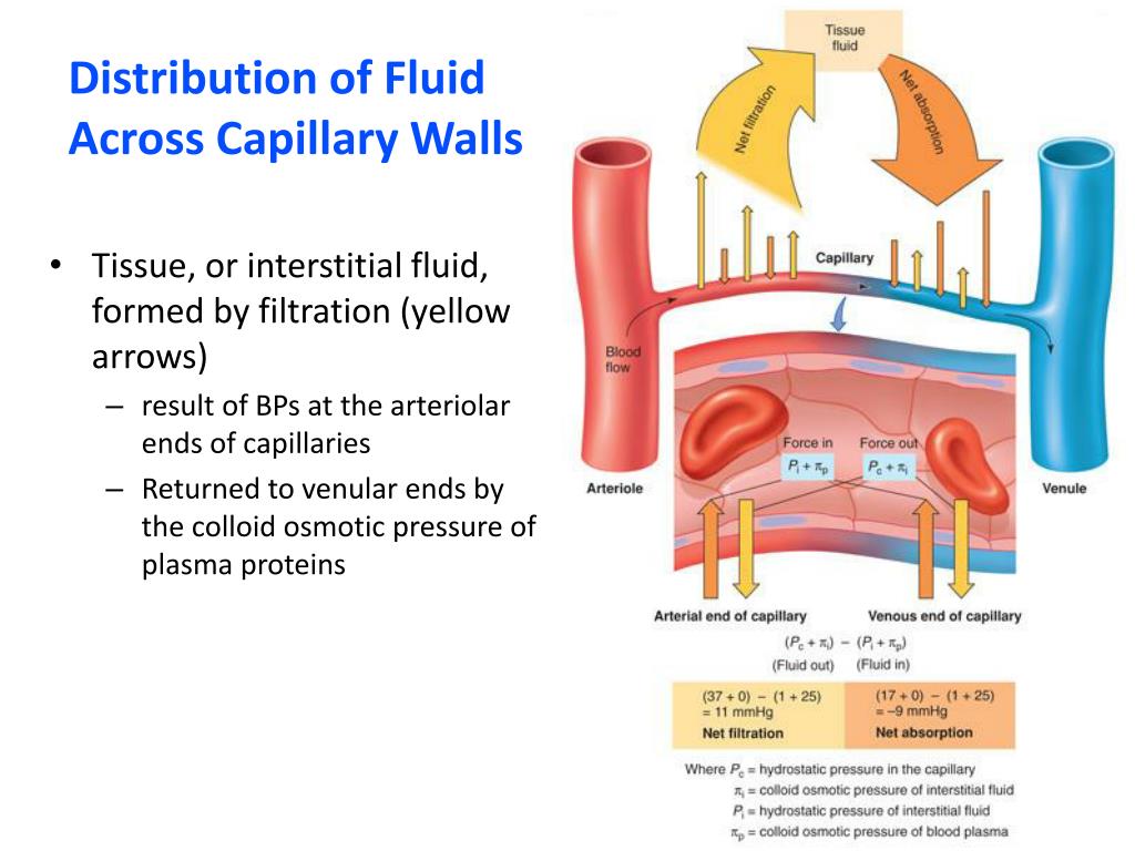
On the arterial side of the capillary, the hydrostatic pressure (the pressure that comes from the heart pumping blood and the elasticity of the arteries) is high. Since capillaries are "leaky" this pressure forces fluid and nutrients against the walls of the capillary and out into the interstitial space and tissues.
How many layers are there in the capillary?
Capillaries are very thin, approximately 5 micrometers in diameter, and are composed of only two layers of cells—an inner layer of endothelial cells and an outer layer of epithelial cells. They are so small that red blood cells need to flow through them single file.
What is the basement membrane?
Surrounding this layer of cells is something called the basement membrane, a layer of protein surrounding the capillary. 2 . If all the capillaries in the human body were lined up in single file, the line would stretch over 100,000 miles.
Where are capillaries found?
Continuous capillaries are generally found in the nervous system, as well as in fat and muscle tissue . Within nervous tissue, the continuous endothelial cells form a blood brain barrier, limiting the movement of cells and large molecules between the blood and the interstitial fluid surrounding the brain.
Where are fenestrated capillaries found?
Fenestrated. These capillaries can be found in tissues where a large amount of molecular exchange occurs, such as the kidneys, endocrine glands, and small intestine. They are particularly important in the glomeruli of the kidneys, as they are involved in filtration of the blood during the formation of urine. The capillaries have small openings in ...
What are the structures that connect the arterioles to the venules?
Capillaries are tiny blood containing structures that connect arterioles to venules. They are small enough to penetrate body tissues, allowing oxygen, nutrients, and waste products to be exchanged between tissues and the blood.
What is sinusoidal capillary?
Sinusoidal capillaries, sometimes referred to as sinusoids, or discontinuous capillaries, have endothelial linings with multiple fenestrations (openings), that are around 30 to 40 nm in diameter. These have no diaphragm and either a discontinuous or non-existent basal lamina.
What is the smallest blood vessel in the body?
Capillaries . Capillaries are tiny blood-containing structures that connect arterioles to venules. They are the smallest and most abundant form of a blood vessel in the body. Capillaries are small enough to penetrate body tissues, allowing oxygen, nutrients, and waste products to be exchanged between tissues and the blood.
Where are sinusoidal capillaries located?
Sinusoidal capillaries are mainly found in the liver, between epithelial cells and hepatocytes. They can also be found in the sinusoids of the spleen where they are involved in the filtration of blood to remove antigens, defective red blood cells, and microorganisms.
What is the function of the precapillary sphincter?
This improves the efficiency of exchange between the blood in the capillary and the tissue surrounding it. Blood flow into the capillaries is controlled by precapillary sphincters, smooth muscle bands that wrap around metarterioles. There are 3 types of capillary in the body; continuous, fenestrated, and sinusoidal.
Where are capillaries located?
A capillary is an extremely small blood vessel located within the tissues of the body that transports blood from arteries to veins. Capillaries are most abundant in tissues and organs that are metabolically active. For example, muscle tissues and the kidneys have a greater amount of capillary networks than do connective tissues .
What are the structures that control the flow of blood through the capillaries?
Microcirculation deals with the circulation of blood from the heart to arteries, to smaller arterioles, to capillaries, to venules, to veins and back to the heart.#N#The flow of blood in the capillaries is controlled by structures called precapillary sphincters. These structures are located between arterioles and capillaries and contain muscle fibers that allow them to contract. When the sphincters are open, blood flows freely to the capillary beds of body tissue. When the sphincters are closed, blood is not allowed to flow through the capillary beds. Fluid exchange between the capillaries and the body tissues takes place at the capillary bed.
What happens to blood pressure in the venule end of the capillary bed?
On the venule end of the capillary bed, blood pressure in the vessel is less than the osmotic pressure of the blood in the vessel . The net result is that fluid, carbon dioxide and wastes are drawn from the body tissue into the capillary vessel.
What controls the flow of blood in the capillaries?
The flow of blood in the capillaries is controlled by structures called precapillary sphincters. These structures are located between arterioles and capillaries and contain muscle fibers that allow them to contract. When the sphincters are open, blood flows freely to the capillary beds of body tissue.
What is the name of the fluids that are exchanged between the blood and the body tissues?
Kes47 / Wikimedia Commons / Public domain. Capillaries are where fluids, gasses, nutrients, and wastes are exchanged between the blood and body tissues by diffusion. Capillary walls contain small pores that allow certain substances to pass into and out of the blood vessel.
What is the net result of fluid moving from the vessel to the body tissue?
The net result is that fluid passes equally between the capillary vessel and the body tissue. Gasses, nutrients, and wastes are also exchanged at this point.
What is the role of capillary walls in the blood pressure?
The capillary walls allow water and small solutes to pass between its pores but does not allow proteins to pass through. As blood enters the capillary bed on the arteriole end, the blood pressure in the capillary vessel is greater than the osmotic pressure of the blood in the vessel.
What is the force that drives fluid out of capillaries and into the tissues?
CHP is the force that drives fluid out of capillaries and into the tissues. As fluid exits a capillary and moves into tissues, the hydrostatic pressure in the interstitial fluid correspondingly rises. This opposing hydrostatic pressure is called the interstitial fluid hydrostatic pressure (IFHP).
What is the primary force that drives fluid transport between the capillaries and tissues?
The primary force driving fluid transport between the capillaries and tissues is hydrostatic pressure, which can be defined as the pressure of any fluid enclosed in a space. Blood hydrostatic pressure is the force exerted by the blood confined within blood vessels or heart chambers. Even more specifically, the pressure exerted by blood against the wall of a capillary is called capillary hydrostatic pressure (CHP), and is the same as capillary blood pressure. CHP is the force that drives fluid out of capillaries and into the tissues.
Why does the CHP dwindle to 18 mm Hg?
Near the venous end of the capillary, the CHP has dwindled to about 18 mm Hg due to loss of fluid. Because the BCOP remains steady at 25 mm Hg, water is drawn into the capillary, that is, reabsorption occurs. Another way of expressing this is to say that at the venous end of the capillary, there is an NFP of −7 mm Hg.
How high is the CHP when blood leaves the arteriole?
When blood leaving an arteriole first enters a capillary bed, the CHP is quite high—about 35 mm Hg. Gradually, this initial CHP declines as the blood moves through the capillary so that by the time the blood has reached the venous end, the CHP has dropped to approximately 18 mm Hg.
What is the function of the lymphatic system?
An important function of the lymphatic system is to return the fluid (lymph) to the blood. Lymph may be thought of as recycled blood plasma. (Seek additional content for more detail on the lymphatic system.) Watch this video to explore capillaries and how they function in the body.
What is the purpose of the cardiovascular system?
The primary purpose of the cardiovascular system is to circulate gases, nutrients, wastes, and other substances to and from the cells of the body. Small molecules, such as gases, lipids, and lipid-soluble molecules, can diffuse directly through the membranes of the endothelial cells of the capillary wall.
What molecules leave the blood through clefts?
Glucose, amino acids, and ions—including sodium, potassium, calcium, and chloride—use transporters to move through specific channels in the membrane by facilitated diffusion. Glucose, ions, and larger molecules may also leave the blood through intercellular clefts.
Which capillaries allow white blood cells to pass through?
However, some capillaries called sinusoidal capillaries (found in the liver, bone marrow and other places) have larger openings between endothelial cells and can allow white and red blood cells to pass through. So the key is: BETWEEN the endothelial cells, not through the cells.
How thick are the walls of the capillary?
The walls of the capillary are only one cell thick ( endothelial cells) - which have spaces between them so stuff from the blood diffuses through the holes in the capillary to the cells beyond the capillary (e.g. liver cells).
What is the capillary made of?
A capillary is made up of a single layer of endothelial cells right. So when blood cells pass through it, do the endothelial cells take blood cells up by endocytosis and then release it to other side. Thanks
Do veins contain blood cells?
So yes, veins connecting capillaries contain blood cells, they just narrow down in size to become capillaries. A capillary is just big enough for a red blood cell to fit through - but they have to go in single file. Nutrients just diffuse through the spaces between the capillary cells.
Do blood cells pass through capillaries?
Normally, blood cells do NOT pass through capillary walls. Capillaries are 'leaky' meaning that plasma and anything dissolved in the plasma will leak through between endothelial cells. Blood cells are just too big to fit. Oxygen, water, and other chemicals pass through the capillary wall.
