What are the sutures of the skull?
Most of the bones of the skull are held together by firm, immovable fibrous joints called sutures or synarthroses. These joints allow the developing skull to grow both pre- and postnatally. The sutures of the skull are morphologically distinct, being divided into three main groups based on the margins of the articulating bones.
What is the difference between a suture and a syndesmoses?
1) Sutures - a fibrous joint between bones of the skull. Functionally, suture joint are a synarthrosis (e.g. sagittal suture, coronal sutures, etc.). They're basically immovable. 2) Syndesmoses - a fibrous joint but there is distance between the articulating bones (e.g. tibiofibular joint). Functionally, they're an ampharthrosis joint.
Is the sagittal suture a synarthrotic joint?
Most of the freely movable joints of the body could be classified both structurally and functionally as. synarthrosis. Because it is an immovable joint, the sagittal suture, which joins the two parietal bones of the skull, is classified as a.
How are the bones of the skull held together?
Most of the bones of the skull are held together by firm, immovable fibrous joints called sutures or synarthroses. These joints allow the developing skull to grow both pre- and postnatally.
What type of joint is a cranial structure?
Suture. All the bones of the skull, except for the mandible, are joined to each other by a fibrous joint called a suture. The fibrous connective tissue found at a suture (“to bind or sew”) strongly unites the adjacent skull bones and thus helps to protect the brain and form the face.
What type of functional joint is a suture?
Three Categories of Functional Joints Synarthrosis: These types of joints are immobile or allow limited mobility. This category includes fibrous joints such as suture joints (found in the cranium) and gomphosis joints (found between teeth and sockets of the maxilla and mandible).
Are cranial sutures cartilaginous joints?
Overview. The joints of the base of the skull are largely cartilaginous joints, or synchondroses. One such joint is the spheno-occipital synchondrosis, which is found between the body of the sphenoid anteriorly and the basilar part of the occipital bone posteriorly.
What is the structural joint category for sutures?
Fibrous joints contain fibrous connective tissue and cannot move; fibrous joints include sutures, syndesmoses, and gomphoses.
What are functional joints?
Functional classifications describe the degree of movement available between the bones, ranging from immobile, to slightly mobile, to freely moveable joints. The amount of movement available at a particular joint of the body is related to the functional requirements for that joint.
Which type of joint includes the sutures of the skull?
fibrous jointA suture is a type of fibrous joint that is only found in the skull (cranial suture). The bones are bound together by Sharpey's fibres. A tiny amount of movement is permitted at sutures, which contributes to the compliance and elasticity of the skull. These joints are synarthroses.
Are sutures fibrous joints?
In fibrous joints the articulating parts are separated by white connective tissue (collagen) fibres, which pass from one part to the other. There are two types of fibrous joints: suture and gomphosis. A suture is formed by the fibrous covering, or periosteum, of two bones passing between them.
What is a primary cartilaginous joint?
Primary cartilaginous joint These cartilaginous joints are composed entirely of hyaline cartilage and are known as synchondroses. Most exist between ossification centres of developing bones and are absent in the mature skeleton, but a few persist in adults.
What are cartilaginous joints?
Cartilaginous joints are where the adjacent bones are joined by cartilage. At a synchondrosis, the bones are united by hyaline cartilage. The epiphyseal plate of growing long bones and the first sternocostal joint that unites the first rib to the sternum are examples of synchondroses.
How are joints classified by both structure and function?
The structural classification divides joints into fibrous, cartilaginous, and synovial joints depending on the material composing the joint and the presence or absence of a cavity in the joint. The functional classification divides joints into three categories: synarthroses, amphiarthroses, and diarthroses.
What is an example of a suture joint?
A suture is the fibrous joint that joins the bones of the skull to each other (except the mandible). A gomphosis is the fibrous joint that anchors each tooth to its bony socket within the upper or lower jaw. The tooth is connected to the bony jaw by periodontal ligaments.
What is a synovial joint?
Synovial joints are the most common type of joint in the body (see image 1). These joints are termed diarthroses, meaning they are freely mobile. A key structural characteristic for a synovial joint that is not seen at fibrous or cartilaginous joints is the presence of a joint cavity.
What is the synovial joint in the skull?
One of them is the atlanto-occipital joint. This is where the condyles on the inferior surface of the occipital bone articulate directly with the C1 vertebra (the atlas ). Flexion and extension of the skull occurs here, allowing the nodding motion of the head as in when we are saying yes.
What is the frontozygomatic suture?
The frontozygomatic suture is a horizontal suture lateral to the orbit. It lies between the frontal process of the zygomatic bone inferiorly and the zygomatic process of the frontal bone superiorly. There is a palpable depression at this suture.
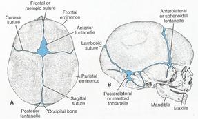
Where is the lambdoid suture located?
The lambdoid suture can be found between the posterior border of the parietal bones and the anterolateral borders of the occipital bone . It is named so due to the lambdoid (λ) shape it forms with the coronal suture. It extends from the posterior extremity of the sagittal suture in a posteroinferior direction to meet the occipitomastoid suture from behind and the parietomastoid suture from above the mastoid process.
Which bone is the articulation of the superior margin of the greater wing of the sphenoid
This suture is another common site for sutural bones to form. The sphenoparietal suture is the articulation of the superior margin of the greater wing of the sphenoid bone and the parietal bone at the angle formed by the convergence of the coronal and squamous sutures at the pterion.
Where are the synchondroses located?
One such joint is the spheno-occipital synchondrosis, which is found between the body of the sphenoid anteriorly and the basilar part of the occipital bone posteriorly.
How many ligaments are there in the temporomandibular joint?
Reinforcing the temporomandibular joint are 3 ligaments. The temporomandibular ligament is a thickening of the lateral aspect of the joint capsule, extending from the zygomatic process and articular ligament of the temporal bone to the neck of the mandible.
What is the second synovial joint?
The second synovial joint is the atlanto-occipital joint, where the base of the skull articulates with the vertebral column. Both these joints will be discussed further in this article.
What is the classification of a joint?
The functional classification of joints is determined by the amount of mobility found between the adjacent bones. Joints are thus functionally classified as a synarthrosis or immobile joint, an amphiarthrosis or slightly moveable joint, or as a diarthrosis, which is a freely moveable joint (arthroun = “to fasten by a joint”). Depending on their location, fibrous joints may be functionally classified as a synarthrosis (immobile joint) or an amphiarthrosis (slightly mobile joint). Cartilaginous joints are also functionally classified as either a synarthrosis or an amphiarthrosis joint. All synovial joints are functionally classified as a diarthrosis joint.
What is an amphiarthrosis joint?
An amphiarthrosis is a joint that has limited mobility. An example of this type of joint is the cartilaginous joint that unites the bodies of adjacent vertebrae. Filling the gap between the vertebrae is a thick pad of fibrocartilage called an intervertebral disc ( [link] ). Each intervertebral disc strongly unites the vertebrae but still allows for a limited amount of movement between them. However, the small movements available between adjacent vertebrae can sum together along the length of the vertebral column to provide for large ranges of body movements.
What is a freely mobile joint?
A freely mobile joint is classified as a diarthrosis. These types of joints include all synovial joints of the body, which provide the majority of body movements. Most diarthrotic joints are found in the appendicular skeleton and thus give the limbs a wide range of motion. These joints are divided into three categories, based on the number of axes of motion provided by each. An axis in anatomy is described as the movements in reference to the three anatomical planes: transverse, frontal, and sagittal. Thus, diarthroses are classified as uniaxial (for movement in one plane), biaxial (for movement in two planes), or multiaxial joints (for movement in all three anatomical planes).