Full Answer
What are the 3 tunics of the eye?
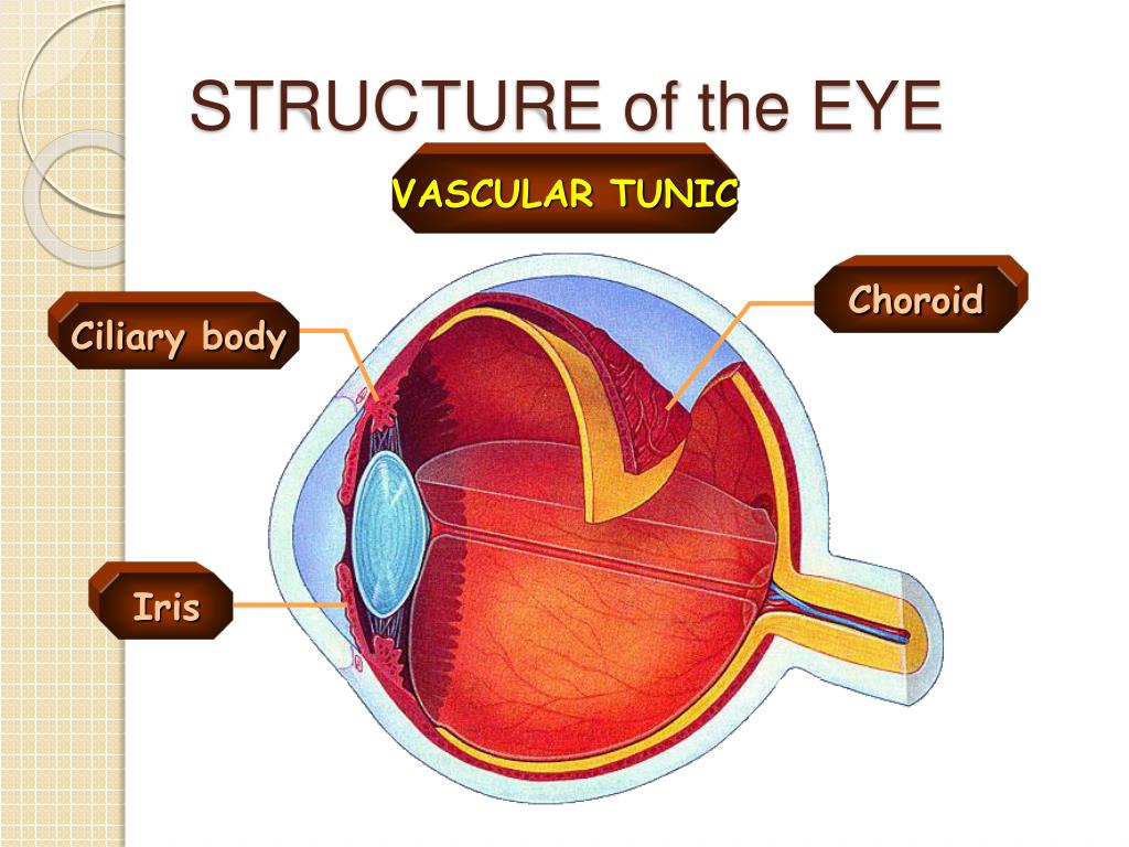
The Tunics of the Eye F IG. 869– Horizontal section of the eyeball. From without inward the three tunics are: (1) A fibrous tunic, (Fig. 869) consisting of the sclera behind and the cornea in front; (2) a vascular pigmented tunic, comprising, from behind forward, the choroid, ciliary body, and iris; and (3) a nervous tunic, the retina.
What is the vascular tunic of the eye?
The Vascular Tunic (tunica vasculosa oculi) (Figs. 872, 873, 874). —The vascular tunic of the eye is formed from behind forward by the choroid, the ciliary body, and the iris. The choroid invests the posterior five-sixths of the bulb, and extends as far forward as the ora serrata of the retina.
What is the projecting transparent part of the external tunic?
The Cornea.—The cornea is the projecting transparent part of the external tunic, and forms the anterior sixth of the surface of the bulb. It is almost circular in outline, occasionally a little broader in the transverse than in the vertical direction.
What makes up the sensory tunic?
The eye is made up of three layers: the outer layer called the fibrous tunic, which consists of the sclera and the cornea; the middle layer responsible for nourishment, called the vascular tunic, which consists of the iris, the choroid, and the ciliary body; and the inner layer of photoreceptors and neurons called the ...May 4, 2010
What is the function of the neural tunic?
The innermost layer of the eye is the neural tunic, or retina, which contains the nervous tissue responsible for photoreception.
What are the 3 tunics of the eye?
The eye itself can be divided into 3 concentric tunics plus the internal components. The three tunics from the outside surface of the eye inward are, (1) the fibrous tunic (cornea and sclera), (2) the vascular tunic (iris, ciliary body, and choroid) and (3) the neuroectodermal (nervous) tunic (retina).
What is a tunic in anatomy?
From Wikipedia, the free encyclopedia. In biology, a tunica (/ˈt(j)uːnɪkə/, UK: /ˈtʃuːnɪkə/; pl. tunicae) is a layer, coat, sheath, or similar covering. The word came to English from the New Latin of science and medicine. Its literal sense is about the same as that of the word tunic, with which it is cognate.
What tunic is the cornea in?
fibrous tunicThe sclera and cornea form the fibrous tunic of the bulb of the eye; the sclera is opaque, and constitutes the posterior five-sixths of the tunic; the cornea is transparent, and forms the anterior sixth.
How many tunics does the eye have?
three tunicsFrom without inward the three tunics are: (1) A fibrous tunic, (Fig. 869) consisting of the sclera behind and the cornea in front; (2) a vascular pigmented tunic, comprising, from behind forward, the choroid, ciliary body, and iris; and (3) a nervous tunic, the retina.
Which structures are part of each tunic?
The vascular tunic is comprised of three distinct regions, (1) the iris, (2) the ciliary body, and (3) the choroid. The vascular tunic is mesodermal in origin and is situated between the outer fibrous tunic and the inner nervous tunic.
Where is aqueous Humour located?
aqueous humour, optically clear, slightly alkaline liquid that occupies the anterior and posterior chambers of the eye (the space in front of the iris and lens and the ringlike space encircling the lens).
What is a tunic in a bulb?
A tunicate bulb has a paper-like covering or tunic that protects the scales from drying and from mechanical injury. Good examples of tunicate bulbs include: tulips, daffodils, hyacinths, grape hyacinths (muscari), and alliums.
What is another word for tunic?
tunicblouse.coat.jacket.robe.chiton.kirtle.surcoat.toga.
What is vascular tunic?
The vascular tunic is comprised of three distinct regions, (1) the iris, (2) the ciliary body, and (3) the choroid. The vascular tunic is mesodermal in origin and is situated between the outer fibrous tunic and the inner nervous tunic. The vascular tunic is also refered to as the uvea.Aug 13, 2020
What are the sensory systems?
A sensory system consists of sensory receptors, neural pathways, and parts of the brain involved in sensory perception. Commonly recognized sensory systems are those for vision, hearing, somatic sensation (touch), taste and olfaction (smell). In short, senses are transducers from the physical world to the realm of the mind.
Which part of the ear is responsible for enhancing sound?
The outer ear collects sound waves in the air and channels them to the inner parts of the ear. The outer ear along with its canal has been shown to enhance sounds within a certain frequency range. That range just happens to be the same range that most of the characteristics of human speech sounds fall into. This allows the sounds to be boosted to twice their original intensity. Parts of the outer ear are the following:
How do tunicates live?
Most tunicates live with the posterior, or lower end of the barrel attached firmly to a fixed object, and have two openings, or siphons, projecting from the other. Tunicates are plankton feeders. They live by drawing seawater through their bodies. Water enters the oral siphon, passes through a sieve-like structure, the branchial basket that traps food particles and oxygen, and is expelled through the atrial siphon.
Do tunicates live alone?
Some kinds of tunicates live alone, and are called solitary tunicates. Others, including the two forms shown here, have the ability to bud off additional individuals from the first to arrive, and these grow into colonies. At first glance, these colonial tunicates look much like other encrusting marine animals, such as sponges. If you look closer, you can see that they have the same structures as solitary tunicates, only much tinier.
What is the sensory modality of touch?
An individual sensory modality represents the sensation of a specific type of stimulus. For example, the general sense of touch, which is known as somatosensation, can be separated into light pressure, deep pressure, vibration, itch, pain, temperature, or hair movement.
What are the sensory receptors?
Sensory Receptors. Stimuli in the environment activate specialized receptors or receptor cells in the peripheral nervous system. Different types of stimuli are sensed by different types of receptors. Receptor cells can be classified into types on the basis of three different criteria: cell type, position, and function.
What is somatosensation in the body?
Somatosensation is the group of sensory modalities that are associated with touch and limb position. These modalities include pressure, vibration, light touch, tickle, itch, temperature, pain, proprioception, and kinesthesia. This means that its receptors are not associated with a specialized organ, but are instead spread throughout the body in a variety of organs. Many of the somatosensory receptors are located in the skin, but receptors are also found in muscles, tendons, joint capsules and ligaments.
What is the role of sensory receptors in the brain?
A major role of sensory receptors is to help us learn about the environment around us, or about the state of our internal environment. Different types of stimuli from varying sources are received and changed into the electrochemical signals of the nervous system. This process is called sensory transduction. This occurs when a stimulus is detected by a receptor which generates a graded potential in a sensory neuron. If strong enough, the graded potential causes the sensory neuron to produce an action potential that is relayed into the central nervous system (CNS), where it is integrated with other sensory information—and sometimes higher cognitive functions—to become a conscious perception of that stimulus. The central integration may then lead to a motor response.
What is the difference between perception and sensation?
Perception is the central processing of sensory stimuli into a meaningful pattern involving awareness. Perception is dependent on sensation , but not all sensation s are perceived. Receptors are the structures (and sometimes whole cells) that detect sensations.
Which sense is most important for autonomic functions?
General senses often contribute to the sense of touch, as described above, or to proprioception (body position) and kinesthesia (body movement), or to a visceral sense, which is most important to autonomic functions.
What is the special sense?
A special sense (discussed in Chapter 15) is one that has a specific organ devoted to it, namely the eye, inner ear, tongue, or nose. Each of the senses is referred to as a sensory modality. Modality refers to the way that information is encoded into a perception.
