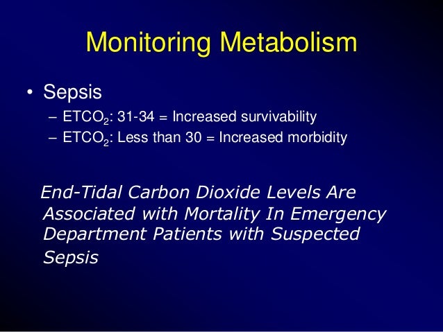
The normal values of ETCO2 is around 5% or 35-37 mm Hg. The gradient between the blood CO2 (PaCO2) and exhaled CO2 (end tidal CO2 or PetCO2) is usually 5-6 mm Hg. PetCO2 can be used to estimate PaCO2 in patients with essentially normal lungs.
What is the normal range of petco2 for COPD?
Most COPD patients we see for CPETs have a PETCO2 of 30 or less and Ve-VCO2 slopes greater than 40. The overall pattern of the patient’s PETCO2 during exercise was also wrong for COPD.
What is the target range for PaCO2 and petco2?
However, the target range for PetCO2 needs to be lower because of the PaCO2-PetCO2 gradient. Where PaCO2 (BGA) is too high, shift the target range 15 mmHg to the left. The %MinVol will increase resulting in a decrease in patient CO2.
What is the normal petco2 level during exercise?
PETCO2 during exercise, a quick diagnostic indicator. The maximum PETCO2 usually occurs at or near anaerobic threshold and the lower limit of normal is around 35 mm Hg. The maximum PETCO2 is reduced below 35 in both cardiac and pulmonary disease and the amount of reduction tends to correlate well with the severity of the disease.
What does a petco2 level of 40 indicate?
The maximum PETCO2 is reduced below 35 in both cardiac and pulmonary disease and the amount of reduction tends to correlate well with the severity of the disease. This by itself is what told me that despite the reduced FEV1 and the diagnosis of COPD, with a PETCO2 of 40 at anaerobic threshold the patient probably had normal gas exchange.
What is a good PetCO2?
With normal physiology, the PaCO2-PetCO2 gradient should be 2-5 mmHg. The acceptable normal ranges for PetCO2 are the same as an arterial blood gas, 35-45 mmHg. If the gradient exceeds 5 mmHg, the clinician should assess for any possible physiologic and equipment factors that may affect the PetCO2 reading.Jul 15, 2021
What does PetCO2 of 8 mm Hg mean?
Waveform capnography PETCO2 levels ≥ 10 mmHg indicate adequate chest compressions. If intra-arterial. relaxation pressure (as measured by using an intra-arterial catheter) during CPR is < 20 mmHg attempt to. improve chest compressions.
What should PetCO2 be during CPR?
Teams should aim for EtCO2 at least >10 mm Hg and ideally >20 mm Hg. Where do these numbers come from? These values are approximately 1/4 the normal EtCO2 (35-45 mm Hg), and ideal CPR will provide at least 1/4 of cardiac output. This is an example of capnography during CPR.Feb 6, 2019
What causes an increase in PetCO2?
Increases in cardiac output and pulmonary blood flow result in better perfusion of the alveoli and a rise in PETCO2.
What does a low PetCO2 mean?
A reduced PetCO2 is often a reflection of gas exchange abnormalities, usually some variation of ventilation-perfusion mismatching.Jun 20, 2013
What is a PetCO2?
Quantitative waveform capnography is the continuous, noninvasive measurement and graphical display of end-tidal carbon dioxide/ETCO2 (also called PetCO2). Capnography uses a sample chamber/sensor placed for optimum evaluation of expired CO2.
What is the main determinant of PETCO2 during CPR?
The main determinant of PETCO2 during CPR is blood delivery to the lungs. Persistently low PETCO2 values less than 10 mm Hg during CPR in intubated patients is a good indicator that achieving ROSC will be unlikely.Dec 25, 2021
When adjusting ventilation rates which PETCO2 value lies within the recommended range?
Avoid excessive ventilation. Ventilation should start at 10/min and should be titrated according to the target PETCO2 of 35-40 mmHg.
What EtCO2 level indicates a problem?
In patients receiving high-quality chest compressions, who have an advanced airway placed, a persistent ETCO2 reading below 10 mm HG after 20 minutes of resuscitation is an indication to terminate efforts [1].Oct 22, 2015
What does an abnormal PETCO2 indicate?
PETCO2 may be a new ventilatory abnormality marker that reflects impaired cardiac output response to exercise in cardiac patients diagnosed with heart failure.
How is PETCO2 calculated?
PACO2 = (K)VCO2/VA (2) An increase in alveolar dead space, which results from the decreased pulmonary vascular pressure, will dilute the CO2 from normally perfused alveolar spaces to decrease PETCO2 below PACO2.
What causes hypercapnia?
Hypercapnia, or hypercarbia, is a condition that arises from having too much carbon dioxide in the blood. It is often caused by hypoventilation or disordered breathing where not enough oxygen enters the lungs and not enough carbon dioxide is emitted.
How does cardiac output affect PETCO 2
Increases in cardiac output and pulmonary blood flow result in better perfusion of the alveoli and a rise in PETCO 2. Under conditions of constant lung ventilation, PETCO 2 monitoring can be used as a monitor of pulmonary blood flow
Cardiac output and (a-ET)PCO 2
Reduction in cardiac output and pulmonary blood flow result in a decrease in PETCO 2 and an increase in (a-ET)PC02.1,2 The percent decrease in PETCO 2 directly correlated with the percent decrease in cardiac output (slope= 0.33, r2=0.82 in 24 patients undergoing aortic aneurysm surgery with constant ventilation).3 Also, the percent decrease in CO 2 elimination correlated with the percent decrease in cardiac output similarly (slope=0.33, r2=0.84).3 The changes in PETCTO2 and CO 2 elimination following hemodynamic perturbation were parallel.
What is the maximum PETCO2 in cardiac patients?
Patients with cardiac disease show a similar pattern to normal patients with the exception that the maximum PETCO2 is reduced below 35 and the degree of reduction correlates well with the NYHA stage of cardiac disease.
How is ETCO2 related to tidal volume?
ETCO2 is related in various degrees to tidal volume, respiratory rate , the deadspace to tidal volume ratio (Vd/Vt) and CO2 production. There is a correlation between PETCO2 and both alveolar CO2 (PACO2) and arterial CO2 (PaCO2) however the correspondance is far from exact or predictable. Alveolar CO2 fluctuates cyclically with ventilation and since Vd/Vt is never zero PETCO2 is always higher than the average PACO2. Numerous investigators have developed algorithms that correlate PETCO2 with arterial CO2 but during exercise PETCO2 can be well below PaCO2 because of ventilatory inefficiency or it increase well above PaCO2 because it can become dominated by mixed-venous PCO2.
Why do COPD patients have mechanical limitations?
COPD patients tend to have both a pulmonary mechanical limitation because of their airway obstruction and a pulmonary vascular (gas exchange) limitation and it’s usually matter of determining which these two factors is the primary limitation.
Is PETCO2 normal during exercise?
The overall pattern of the patient’s PETCO2 during exercise was also wrong for COPD. The PETCO2 pattern that normal patients show during a CPET is to start off with a relatively low PETCO2. The PETCO2 then increases to its maximum value (usually at AT) and then decreases to peak exercise.
Does PETCO2 rise with COPD?
COPD patients also tend to have distinct pattern, that is pretty much the opposite of pulmonary hypertension. For COPD patients PETCO2 tends to rise throughout testing. Again if there is an anaerobic threshold, the PETCO2 at that time also tends not to be the maximum PETCO2.
Does PETCO2 decrease with pulmonary hypertension?
Patients with pulmonary hypertension however, show a distinctly different pattern where PETCO2 declines throughout testing and if anaerobic threshold is attained, the PETCO2 at that time is not the maximum PETCO2. COPD patients also tend to have distinct pattern, that is pretty much the opposite of pulmonary hypertension.
Is a PETCO2 of 40 normal?
This by itself is what told me that despite the reduced FEV1 and the diagnosis of COPD, with a PETCO2 of 40 at anaerobic threshold the patient probably had normal gas exchange. We do not routinely do diffusion capacity testing as part of a cardiopulmonary exercise test so that part will have to remain speculative. When the Ve-VCO2 slope was calculated however, it was 28, which is well within normal limits. Most COPD patients we see for CPETs have a PETCO2 of 30 or less and Ve-VCO2 slopes greater than 40.
Why is the target range for PetCO2 lower?
However, the target range for PetCO2 needs to be lower because of the PaCO2-PetCO2 gradient.
When should you record petCO2?
You should record the measured patient PetCO2 value just before the BGA.
Is PaCO2 higher than PetCO2?
Under common conditions, PaCO2 is approximately 3–5 mmHg higher than PetCO2 — the difference between the values is referred to as the PaCO2-PetCO2 gradient. Under special clinical conditions (including ventilation/perfusion problems or presence of a shunt), the PaCO2-PetCO2 gradient can increase, requiring adjustment of the ventilation targets. The Target Shift control (INTELLiVENT-ASV window) allows you to move the PetCO2 target range to the left (lower values) or to the right (higher values) within the limits defined.

Some Physiology
- Also, according to the AHA, continuous waveform capnography along with clinical assessment is the most reliable method of confirming and monitoring correct placement of an ET tube.
Some History of Old Fashioned Monitors
Pulse Oximetry
End Tidal CO2 Has Many Uses