What role does the ligamentum arteriosum play in major trauma?
The ligamentum arteriosum plays a role in major trauma. It fixes the aorta in place during abrupt motions, consequently potentially resulting in a ruptured aorta.
What is the function of the ligamentum arteriosum in larynx?
After splitting from the left vagus nerve, the left recurrent laryngeal loops around the aortic arch behind the ligamentum arteriosum, after which it ascends to the larynx. In adults, the ligamentum arteriosum has no useful function.
Why does the ductus arteriosus become the ligamentum arteriosum?
This significantly reduces the volume of blood ciruclating through the lungs, which are inactive in the womb. The ductus arteriosus becomes the ligamentum arteriosum within three weeks of birth, so that deoxygenated blood can be selectively circulated to the lungs for more efficient oxygenation of the blood.
Why does the ligament arteriosum keep the aorta in place?
These abrupt rate would cause the heart to launch forward, but since the ligament arteriosum is keeping the aorta in place, it would prevent the dominant force from detaching and tearing the aorta completely.
What is the function of the ductus arteriosus?
The ductus arteriosus is a normal fetal artery connecting the aorta and the main lung artery (pulmonary artery). The ductus allows blood to detour away from the lungs before birth. Every baby is born with a ductus arteriosus.
What vessels does the ligamentum arteriosum connect?
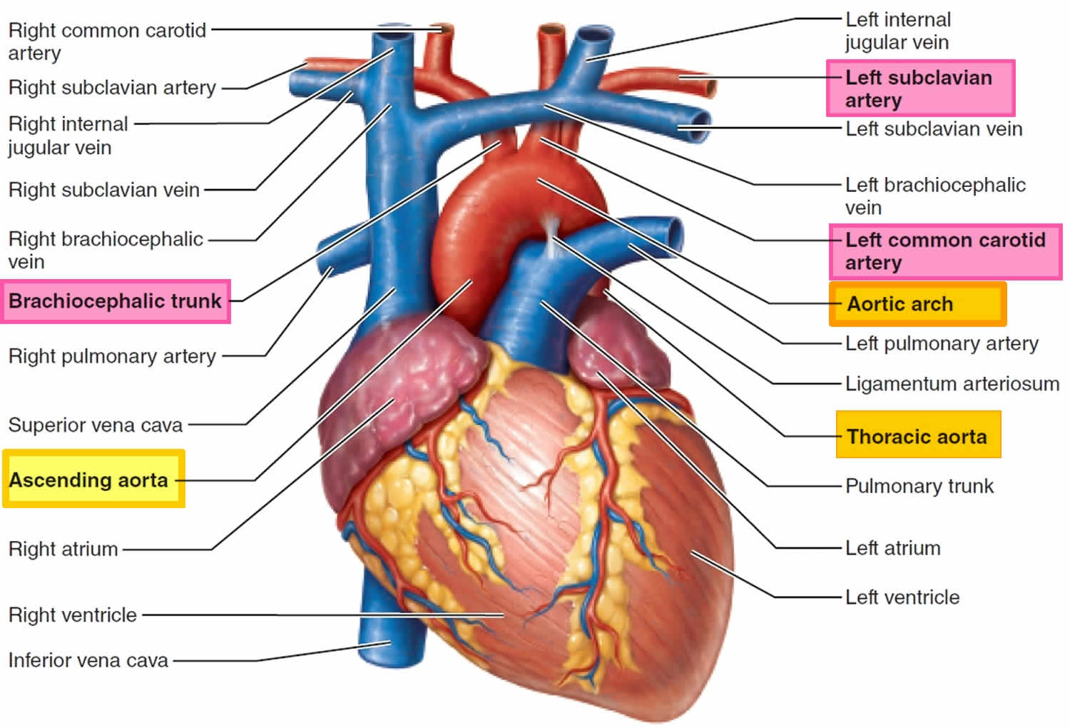
The ligamentum arteriosum (or arteriosus) is the small fibrous remnant of the fetal ductus arteriosum, located between and connecting the proximal left pulmonary artery and the undersurface of the junction of the aortic arch and descending aorta, at the aortic isthmus.
What is the difference between the ligamentum arteriosum and the ductus arteriosus?
Ligamentum arteriosum (also known as Ligament of Botallo or Harvey's ligament) is a fibrous remnant of the fetal ductus arteriosus (ductus Botalli, Botallo's duct). The ductus arteriosus is a vessel connecting the pulmonary trunk and the aortic arch or descending aorta in the fetus.
How is ligamentum arteriosum formed?
Introduction. The ligamentum arteriosum is generally considered to be a mere a remnant of the embryonic bypass (ductus arteriosus) from the pulmonary circulation to the aortic arch, obliterating soon after childbirth.
What is the developmental significance of the ligamentum arteriosum and Fossa Ovalis?
The ligamentum arteriosum is the vestigial structure that is seen in adults. Instead of connecting the pulmonary arteries and aorta, it helps to stabilize these structures. The foramen ovale is a fetal window between the right and left atria to help blood bypass the pulmonary circuit.
What keeps ductus arteriosus open?
Prostaglandin E1 (PGE1) is a substance produced by the ductus that keeps it open. External PGE1 is used to keep the ductus arteriosus open in neonates who have heart lesions that depend on an open ductus for survival.
What is the function of the ductus arteriosus quizlet?
In the developing fetus, the ductus arteriosus, is a blood vessel connecting the pulmonary artery to the aortic arch. It allows most of the blood from the right ventricle to bypass the fetus's fluid-filled non-functioning lungs. Upon closure at birth, it becomes the ligamentum arteriosum.
What is the function of ductus arteriosus during fetal life?
The ductus arteriosus sends the oxygen poor blood to the organs in the lower half of the fetal body. This also allows for the oxygen poor blood to leave the fetus through the umbilical arteries and get back to the placenta to pick up oxygen.
What is the ductus arteriosus called after birth?
Patent ductus arteriosus (PDA) is a persistent opening between the two major blood vessels leading from the heart. The opening (ductus arteriosus) is a normal part of a baby's circulatory system in the womb that usually closes shortly after birth. If it remains open, it's called a patent ductus arteriosus.
What is the relationship between the ductus arteriosus and the ligamentum arteriosum?
The ductus arteriosus responds to these changes by closing and becoming the ligamentum arteriosum. This prevents oxygenated blood from returning to the pulmonary circulation and after passing through the lungs and into the aorta. This closure of the ductus occurs in most individuals within the first 3 months of life.
What are the coronary vessels?
Coronary arteries are the blood vessels that supply oxygen-rich blood to your heart muscle to keep it pumping. The coronary arteries are directly on top of your heart muscle.
Which vessel is part of the coronary circulation?
Coronary circulation is the circulation of blood in the blood vessels that supply the heart muscle (myocardium). Coronary arteries supply oxygenated blood to the heart muscle. Cardiac veins then drain away the blood after it has been deoxygenated....Coronary circulationMeSHD003326Anatomical terminology2 more rows
What are subclavian vessels?
The subclavian artery is a paired blood vessel that provides blood supply to the upper limbs, as well as parts of the neck and brain.
What is the ductus arteriosus?
The ductus arteriosus is a vessel connecting the pulmonary trunk and the aortic arch in the fetus. While this is a vestigial structure in an adult, during fetal development, the ductus arteriosus’ function is to bypass the lungs. Normally, the ductus arteriosus closes soon after birth; however, occasionally it may remain open, ...
What is the ligament of the fetal ductus?
Ligamentum arteriosum and ductus arteriosus. Ligamentum arteriosum (also known as Ligament of Botallo or Harvey's ligament) is a fibrous remnant of the fetal ductus arteriosus (ductus Botalli, Botallo's duct). The ductus arteriosus is a vessel connecting the pulmonary trunk and the aortic arch in the fetus. While this is a vestigial structure in an ...
What is PDA in medical terms?
Patent ductus arteriosus (PDA) The closure of the ductus arteriosus is controlled by a number of factors and if one or more of them become disrupted, the closure may not occur, resulting in a condition known as patent ductus arteriosus.
How long does it take for blood flow to reverse in the ductus arteriosus?
Although some degree of reversed blood flow may be present in the ductus arteriosus for up to a week, eventually, complete closure and breakdown occurs, leaving the fibrous structure we observe in adults, known as ligamentum arteriosum.
How long does it take for anatomic closure to occur?
Anatomic closure occurs gradually in the following 1-3 months by the means of thickening of the tunica intima, the innermost layer of the vessel. In short, this means that no more blood is physically able to enter the ductus arteriosus and it begins to gradually break down.
What receptors are inhibited by prostaglandins?
However, in an unborn fetus, these receptors are inhibited by prostaglandins E2, I2 and F2a, ...
Which aortic arch is the precursor of the ductus arteriosus?
Initially, the recurrent laryngeal branches on both sides loop under the 6th aortic arch, the precursor of the ductus arteriosus. The 6th aortic arch past the right pulmonary artery atrophies, causing the right recurrent laryngeal branch to ascend to the right subclavian artery, while on the left, the ductus arteriosus anchors it inferior to ...
How does the ductus arteriosus affect the lungs?
The ductus arteriosus becomes the ligamentum arteriosum within three weeks of birth, so that deoxygenated blood can be selectively circulated to the lungs for more efficient oxygenation of the blood.
What is the ligamentum arteriosum?
Left side. (Lig. arteriosum labeled at upper right.) Heart of dog. The ligamentum arteriosum ( Latin: arterial ligament) is a small ligament that is the remnant of the ductus arteriosus formed within three weeks after birth .
What happens if the ductus arteriosus fails to close?
Such ruptures are very rare. If the ductus arteriosus fails to close after birth, a condition known as patent ductus arteriosus can develop. This is a fairly common birth defect. Sufferers may have operations that leave them with no ligamentum arteriosum.
Which artery is ligamentum arteriosum attached to?
On the other, inferior end, the ligamentum is attached to the top of the left pulmonary artery. The ligamentum arteriosum is closely related to the left recurrent laryngeal nerve, a branch of the left vagus nerve.
What is a coarctation of the aorta?
Coarctation of the aorta (CoA) refers to a narrowing in the descending aorta ( Figure 74-1) , typically in the location of the takeoff of the left subclavian artery and the ligamentum arteriosum (ductus arteriosus). Anatomically it can occur just before the ductus, at the ductus, or just after the ductus. In its rarest form, CoA can involve the abdominal segment of the aorta (<2% of all cases).1 Adults with a previously undiagnosed CoA will almost always have postductal lesions. The lesion itself is either a localized narrowing or a hypoplastic segment of the aorta, with the former lesion being much more common. CoA is a very common defect, accounting for an estimated 8% of all congenital heart defects. 2 Although distributed fairly evenly between genders among infants, older patients have a male predominance. Approximately 50% to 85% of patients with CoA also have a bicuspid aortic valve. 3 If there are no other anatomic abnormalities (with the exception of the biscupid valve), the lesion is referred to as a “simple coarctation.” 4 If accompanied by other cardiac anomalies it is referred to as a “complex coarctation.”
What causes constriction of the esophagus?
Persistence of the right fourth aortic arch causes constriction of the esophagus with dysphagia and regurgitation. The aorta is situated to the right of the esophagus and trachea and the ligamentum arteriosum in its connection to the pulmonary artery encloses the esophagus in a vascular ring and compresses it against the trachea.
What is the definition of a congenital abnormality in which the right fourth aortic arch becomes the
Definition. Rare congenital abnormality in which the right fourth aortic arch becomes the definitive aorta instead of the left aortic arch. The esophagus becomes constricted by the ligamentum arteriosum as it extends between the anomalous right aorta and the left pulmonary artery.
What is the coarctation of the thoracic spinal cord?
With regards to steal, the upper thoracic spinal cord segment is a watershed zone of spinal perfusion.
Where is the recurrent laryngeal nerve located?
The recurrent laryngeal nerve (RLN) is derived from the vagus trunk as it enters the thorax. The left RLN curves below and behind the aortic arch at the level of the ligamentum arteriosum. The right RLN loops under the subclavian artery. Then both nerves ascend lateral to the trachea, run in the tracheoesophageal groove and behind the thyroid, toward the larynx. The RLNs provide ipsilateral motor innervation to the intrinsic laryngeal muscles for vocalization—to all the intrinsic laryngeal muscles except for the cricothyroids. These muscles are responsible for the abduction and adduction of the vocal cords, thereby playing a major role in speech, swallowing, and breathing (Krasna & Forti, 2006 ). The nerves also supply motor innervation to the inferior pharyngeal constrictor muscles. As for their sensory component, they provide sensory innervation to the upper esophagus and trachea and mediate airway sensation from the level of the true vocal cords to the carina ( Aquino, Duncan, & Hayman, 2001 ). They also contain autonomic parasympathetic fibers that are given off as cardiac branches supplying the cardiac plexus.
Which aorta is constricted by ligamentum arteriosum?
Congenital abnormality in which the right fourth aortic arch becomes the definitive aorta instead of the left aortic arch. The esophagus becomes constricted by the ligamentum arteriosum as it extends between the anomalous right aorta and the left pulmonary artery.
Where is the most common manifestation of a ductus arteriosus?
The most common manifestation is immediately distal to the ligamentum arteriosum. Pathologically, it is thought to develop from the same process that obliterates the ductus arteriosus. It is hypothesized that oxygen-sensitive smooth muscle tissue from the ductus becomes incorporated within the aortic wall.
What is ligamentum in Ludwig?
The ligamentum may create a small diverticulum arising from the anteromedial wall of the aorta , known as a ductus diverticulum, that may be mistaken for a aneurysm.
Where is the ligamentum arteriosum located?
The ligamentum arteriosum (or arteriosus) is the small fibrous remnant of the fetal ductus arteriosum, located between and connecting the proximal left pulmonary artery and the undersurface of the junction of the aortic arch and descending aorta, at the aortic isthmus. The left recurrent laryngeal nerve curves upwards ...