What is the function of the fovea centralis Quizlet?
The function of the fovea centralis is to allow for sharp and focused vision. It is needed for activities such as reading, driving, knitting, etc. Why does the fovea have the highest acuity?
What is the fovea centralis made of?
Fovea centralis. The fovea centralis is a small, central pit composed of closely packed cones in the eye. It is located in the center of the macula lutea of the retina.
What is the difference between the macula and fovea centralis?
The macula contains mostly cones and few rods, and the fovea centralis contains only cones and no rods. In the eye disease known as age-related macular degeneration, or AMD, the cones are damaged by a buildup of toxic products of eye metabolism called drusin.
What is the function of the fovea?
The fovea or fovea centralis is a small depression at the center of the retina that's responsible for central vision. It's the point at which visual acuity is at its highest. Visual acuity is the ability to identify the details of objects when you look at them.
What is the main function of the fovea centralis?
The fovea is responsible for sharp central vision (also called foveal vision), which is necessary in humans for reading, driving, and any activity where visual detail is of primary importance.
What is the structure and function of the fovea?
A fovea is a pitted invagination in the inner retina that overlies an area of densely packed photoreceptors specialized for high acuity vision. A fovea contains particularly high numbers of photoreceptors and neurons, and provides the highest visual resolution (Walls, 1942).
What is the meaning of fovea centralis?
fovea centralis. [ sĕn-trā′lĭs ] n. A depression in the center of the macula of the retina, the area of the most acute vision, where only cones are present and where blood vessels are lacking. central fovea central pit.
How is fovea centralis adapted to its function?
Cones in the central fovea express pigments that are sensitive to green and red light. These cones are the 'midget' pathways that also underpin high acuity functions of the fovea. The fovea is employed for accurate vision in the direction where it is pointed.
What is the fovea centralis quizlet?
The fovea centralis is a small, central pit composed of closely packed cones in the eye. It is located in the center of the macula lutea of the retina. The fovea is responsible for sharp central vision such as reading and driving.
Where is the fovea centralis located in the eye and what is the function of the fovea centralis?
The fovea centralis is an indented area located in the center of the macula of the retina. The cells of the retina are rods and cones. Rods play a role in night vision, while cones enable central, daytime vision. The fovea centralis consists of only cones and is, therefore, crucial to central vision.
What is macula and fovea centralis?
The macula is the center portion of the retina that produces even sharper vision with its rods and cones. The fovea is the pit inside the macula with only cones, so vision can be at its sharpest. While the fovea and the macula have the same objective of providing clear vision, they achieve that goal in different ways.
Is the fovea responsible for central vision?
The fovea centralis, or fovea, is a small depression within the neurosensory retina where visual acuity is the highest. The fovea itself is the central portion of the macula, which is responsible for central vision.
Why is the fovea centralis the area of the sharpest vision?
The fovea has the densest concentration of photoreceptor cells that are known as cones. Rods are completely absent from the fovea. The ganglion and bipolar layers of the retina spread apart at the fovea to give light a direct path to the cones for the sharpest vision.
Is the fovea centralis a blind spot?
The blind spot (Fovea centralis) This seemingly poor design of the retina, which produces the blind spot in our visual field, is referred to by experts as the inverted eye. The blind spot is located about 15 degrees on the nasal side of the fovea.
Is the fovea the blind spot?
fovea, the blind spot. The area where the optic nerve connects to the retina in the back of each eye is known as the optic disk. There is a total absence of cones and rods in this area, and, consequently, each eye is completely blind in this spot.
What differentiates the fovea centralis from the rest of the retina?
The foveal center or 'foveola' contains the highest density of cone photoreceptors in the retina. Cone photoreceptors function in bright light and support high acuity and color vision.
What is the difference between macula lutea and Fovea Centralis?
Fovea centralis is a part of the macula lutea. It is a small depression within the central part of the macula lutea that allows for sharp vision.
What is the function of the macula lutea?
The function of the macula lutea is to allow for sharp and focused vision in the human eye. It is a yellow spot in the center of the retina.
What is the function of the fovea?
The function of the fovea centralis is to allow for sharp and focused vision. It is needed for activities such as reading, driving, knitting, etc.
Why does the fovea have the highest acuity?
The fovea centralis has the highest visual acuity of the entire retina because it consists of a high density of cones. Cones are responsible for co...
What is fovea made up of?
The fovea centralis is made up entirely of cones. Cones are a type of photoreceptor that allows for sharp vision and visual acuity.
Anatomy of the Eye
The human body consists of five important senses: vision, smell, taste, hearing, and touch. In humans, the sense of sight relies on the eyes as its primary organs. The human eye consists of many different anatomical structures that help the eye achieve its function.
What is the Fovea Centralis?
This section will answer the question of what is the fovea centralis. The term 'fovea' means a small fossa, which is a cavity or depression, whereas the term 'centralis' means central or in the middle in Latin. Thus, the term fovea centralis translates to a small fossa in the middle.
Fovea Centralis Structure
The fovea centralis is a small depression in the retina that measures about 1.5mm in diameter. It is shaped like a horizontal elliptical disc. There is a 0.5mm wide area within the fovea centralis that is called the foveal avascular zone. This area does not contain any blood vessels.
What is the fovea centralis?
Fovea centralis. The fovea is a tiny part of the eye’s anatomy that makes a huge difference in our eyesight. Resting inside the macula, the fovea (also called “fovea centralis”) provides our absolute sharpest vision.
Why is the fovea important?
Because the fovea is such an essential part of a person’s vision, it’s important to prevent and/or monitor the conditions that may jeopardize its function. Conditions that may affect the fovea include:
Why is the fovea anatomy so tricky?
Fovea anatomy can be tricky because the retina and macula are also light-sensitive parts of the eye that create sharp vision. So, where does the fovea come into play, and how is it different from the macula and retina?
Which part of the retina produces sharper vision?
The macula is the center portion of the retina that produces even sharper vision with its rods and cones. The fovea is the pit inside the macula with only cones, so vision can be at its sharpest. While the fovea and the macula have the same objective of providing clear vision, they achieve that goal in different ways.
How to protect your eyes from fovea?
The best way to protect your sight and keep your eyes healthy is to undergo routine comprehensive eye exams — it may be possible for an eye doctor to detect early signs of a fovea-related condition before you notice any symptoms.
Where does the name Fovea come from?
The name “fovea” comes from the Greek word meaning “small pit.”. This is an appropriate name, as the fovea is a tiny depression (or pit) in the macula, a small structure located in the center of the retina, the light-sensitive tissue that lines the back of the eye.
What are cone cells responsible for?
Cone cells are responsible for producing color and fine details , while rods provide peripheral vision, movement and shades of grey. Rods are mostly located outside the macula, and the cones are located inside. The fovea eye pit does not have any rods or other neurons, only millions of tightly packed cones.
Anatomy
The central fovea appears as a small flat spot at the retina's center. It's about 1.5 mm in diameter and contains about 199,000 cones/mm squared. 2
Fovea Problems & Diseases
Left untreated, a variety of eye diseases may impair the fovea, resulting in vision loss.
Why Routine Eye Exams are Important
Routine treatment is important because it helps your eye doctor detect any issues before they become serious.
What is the central fovea?
The central fovea consists of very compact cones, thinner and more rod-like in appearance than cones elsewhere. These cones are very densely packed (in a hexagonal pattern). Starting at the outskirts of the fovea, however, rods gradually appear, and the absolute density of cone receptors progressively decreases.
What is the fovea?
The fovea is responsible for sharp central vision (also called foveal vision), which is necessary in humans for activities for which visual detail is of primary importance , such as reading and driving. The fovea is surrounded by the parafovea belt and the perifovea outer region.
How do pigments enhance the acuity of the fovea?
The pigments also enhance the acuity of the fovea by reducing the sensitivity of the fovea to short wavelengths and counteracting the effect of chromatic aberration. This is also accompanied by a lower density of blue cones at the center of the fovea. The maximum density of blue cones occurs in a ring about the fovea.
What is the size of the fovea?
Structure. The fovea is a depression in the inner retinal surface, about 1.5 mm wide, the photoreceptor layer of which is entirely cones and which is specialized for maximum visual acuity. Within the fovea is a region of 0.5mm diameter called the foveal avascular zone (an area without any blood vessels).
How far is the Perifovea from the Fovea?
The parafovea extends to a radius of 1.25 mm from the central fovea, and the perifovea is found at a 2.75 mm radius from the fovea centralis. The term fovea comes from the from Latin foves 'pit'.
Where is the fovea found?
Other animals. The fovea is also a pit in the surface of the retinas of many types of fish, reptiles, and birds. Among mammals, it is found only in simian primates. The retinal fovea takes slightly different forms in different types of animals.
What is the optic disc in a fundus photograph?
A fundus photograph showing the macula as a spot to the left. The optic disc is the area on the right where blood vessels converge. The grey, more diffuse spot in the centre is a shadow artifact .
Anatomy
Functions
- The fovea enables sharp central vision (foveal vision). This type of vision enables you to perform activities that require visual detail like reading, writing, or driving. To see well, you must focus the image on the fovea centralis. This is the reason why you unconsciously move your head or eyes when reading this sentence — to center the words on ...
Fovea Problems & Diseases
- Left untreated, a variety of eye diseases may impair the fovea, resulting in vision loss. The following are examples of fovea-related issues:
Why Routine Eye Exams Are Important
- Routine treatment is important because it helps your eye doctor detect any issues before they become serious. During a routine eye exam, your opthamologist will check for all common eye issues such as cataracts, macular degeneration, tears, or detachments. They'll also give you advice on best care practices to keep your eyes healthy.
Other Parts of The Eye
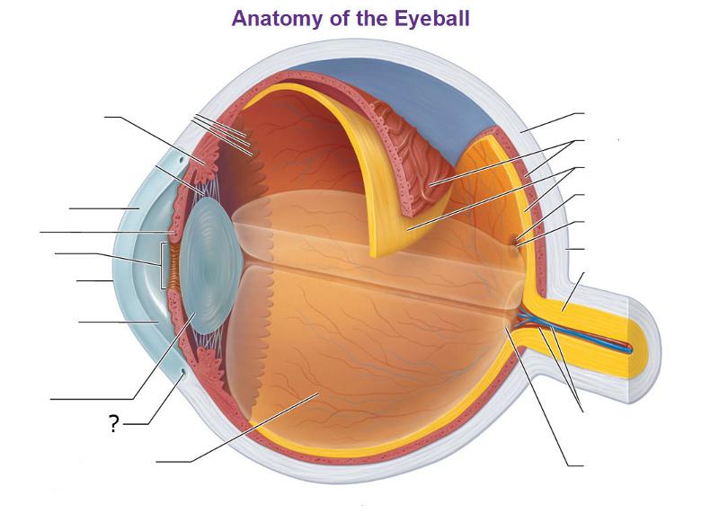
- Other important parts of the eye include: 1. The cornea. The clear bulging surface of the eye. 2. Sclera. The white part of the eye. 3. Iris.The color part that surrounds the pupil. 4. The lens of the eye. The clear part of the eye that helps with focus. 5. Pupil. An opening through which light passes into the eye. 6. Vitreous Humor. The fluid-filled space between the lens and the retina.
Overview
The fovea centralis is a small, central pit composed of closely packed cones in the eye. It is located in the center of the macula lutea of the retina.
The fovea is responsible for sharp central vision (also called foveal vision), which is necessary in humans for activities for which visual detail is of primary importance, such as reading and driving. The fovea is surrounded by the parafovea belt and the perifovea outer region.
Function
In the primate fovea (including humans) the ratios of ganglion cells to photoreceptors is about 2.5; almost every ganglion cell receives data from a single cone, and each cone feeds onto between one and 3 ganglion cells. Therefore, the acuity of foveal vision is limited only by the density of the cone mosaic, and the fovea is the area of the eye with the highest sensitivity to fine details. Cones in the central fovea express pigments that are sensitive to green and red light. These cones are t…
Structure
The fovea is a depression in the inner retinal surface, about 1.5 mm wide, the photoreceptor layer of which is entirely cones and which is specialized for maximum visual acuity. Within the fovea is a region of 0.5mm diameter called the foveal avascular zone (an area without any blood vessels). This allows the light to be sensed without any dispersion or loss. This anatomy is responsible for the depression in the center of the fovea. The foveal pit is surrounded by the foveal rim that cont…
Other animals
The fovea is also a pit in the surface of the retinas of many types of fish, reptiles, and birds. Among mammals, it is found only in simian primates. The retinal fovea takes slightly different forms in different types of animals. For example, in primates, cone photoreceptors line the base of the foveal pit, the cells that elsewhere in the retina form more superficial layers having been displaced away from the foveal region during late fetal and early postnatal life. Other foveae may s…
Additional images
• Illustration showing main structures of the eye including the fovea
• Structures of the eye labeled
• This image shows another labeled view of the structures of the eye
• Schematic diagram of the macula lutea of the retina, showing perifovea, parafovea, fovea, and clinical macula
See also
• Eye movement
• Gaze-contingency paradigm
• Macular degeneration
• Foveated imaging