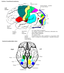
What is elevated portions of the cerebral cortex?
| Question | Answer |
| elevated portions of the cerebral cortex ... | Gyri |
| causalgia | burning sensation of pain |
| a network of interlacing nerve fibers in ... | Plexus |
What is the medical term meaning elevated portion of the cerebral cortex?
- Answers What is the medical term meaning Elevated portion of the cerebral cortex? A Ridge/Hill is called a Gyrus (pl Gyri) and a cleft/valley is called a Sulcus (pl Sulci). Some of the Gyri and Sulci are individually named for example look at the cerebrum from a lateral view.
What are the peaks and troughs of the cerebral cortex?
Most mammals have a cerebral cortex that is convoluted with the peaks known as gyri and the troughs or grooves known as sulci. Some small mammals including some small rodents have smooth cerebral surfaces without gyrification. The larger sulci and gyri mark the divisions of the cortex of the cerebrum into the lobes of the brain.
What are the association areas of the cerebral cortex?
The association areas are the parts of the cerebral cortex that do not belong to the primary regions. They function to produce a meaningful perceptual experience of the world, enable us to interact effectively, and support abstract thinking and language.
What is the thickness of the cerebral cortex?
The thickness of the cerebral cortexvaries from 2 to 6 mm. In higher mammals likehumans, the cerebral cortex looks likeit has many bumps and grooves. A bump or bulge on the cortexis called a gyrus (the plural of the word gyrus is "gyri") and a groove is called a sulcus (the plural of the word sulcus is "sulci").
What are the raised portions of the brain?
Gyri (singular: gyrus) are the folds or bumps in the brain and sulci (singular: sulcus) are the indentations or grooves. Folding of the cerebral cortex creates gyri and sulci which separate brain regions and increase the brain's surface area and cognitive ability.
What does the cerebral cortex do for the brain?
Each of these lobes is responsible for processing different types of information. Collectively, your cerebral cortex is responsible for the higher-level processes of the human brain, including language, memory, reasoning, thought, learning, decision-making, emotion, intelligence and personality.
What happens when the cerebral cortex is damaged?
The cerebral cortex plays a crucial role in nearly all brain functions. Damage to it can cause many cognitive, sensory, and emotional difficulties.
What gives rise to cerebral cortex?
The medial ganglionic eminence gives rise to a population of early neurons in the developing cerebral cortex.
What does cortex mean in medical terms?
Medical Definition of cortex 1a : the outer or superficial part of an organ or body structure (as the kidney, adrenal gland, or a hair) especially : cerebral cortex. b : the outer part of some organisms (as paramecia)
What part of the brain is the cerebral cortex?
cerebrumThe cerebral cortex is the thin layer of the brain that covers the outer portion (1.5mm to 5mm) of the cerebrum. It is covered by the meninges and often referred to as gray matter. The cortex is gray because nerves in this area lack the insulation that makes most other parts of the brain appear to be white.
Can a person survive without a cerebral cortex?
There are a surprising number of known cases of people missing half of their cerebral cortex—the outermost chunk of brain tissue. A currently living and healthy 16-year-old German girl is one. She was born without the right hemisphere of her cortex, though this wasn't discovered until she was 3 years old.
Does brain damage always show on MRI?
And the answer is if it's moderate or severe, most of the time it will show up on an MRI. If it's a mild brain injury, often it will not show up on an MRI.
Can the brain heal itself from brain damage?
And the answer is yes. The brain is incredibly resilient and possesses the ability to repair itself through the process of neuroplasticity. This phenomenon is the reason why many brain injury survivors can make astounding recoveries.
What age does the cerebral cortex develop?
The formation of the cortex begins with the appearance of the neural plate at around 18 days of gestation. Two days later, one can distinguish the three major divisions of the brain. Differentiation of the cerebral vesicles occurs at about day 33 of gestation.
How does alcohol affect the cerebral cortex?
In the cerebral cortex, alcohol can a ect thought processes, leading to potentially poor judgment. Alcohol depresses inhibition, leading one to become more talkative and more confident. Alcohol blunts the senses and increases the threshold for pain.
Is the cerebral cortex the same as the cerebrum?
The main difference between cerebrum and cerebral cortex is that cerebrum is the largest part of the brain whereas cerebral cortex is the outer layer of the cerebrum. The cerebrum comprises two cerebral hemispheres. The cerebral cortex is made up of gray matter that covers the internal white matter.
What is the largest part of the brain?
The cerebrum, also known as the forebrain, is the largest part of the brain. It is derived embryologically from the telencephalon. The cerebrum consists of two cerebral hemispheres (right and left) separated by a deep longitudinal fissure which contains the corpus callosum.
Which lobe of the cerebrum is the most anterior?
The frontal lobe is the most anterior part of the cerebrum. It is involved in activities like muscle control, higher intellect, personality, mood, social behaviour, and language. Posteriorly, the frontal lobe is separated from the parietal lobe by the central sulcus (of Rolando) and inferiorly from the temporal lobe by the lateral sulcus (of Sylvius). The most significant convolutions of the frontal lobe are the precentral, superior, middle, inferior and orbital gyri. The entire frontal lobe is supplied by the anterior and middle cerebral arteries, which are branches of the internal carotid artery.
What is the parietal lobe?
The parietal lobe is situated between the frontal and occipital lobes, and separated from them by the central and parieto-occipital sulci respectively. It is involved in language and calculation, as well as the perception of various sensations such as touch, pain, and pressure. The lobe consists of two parts called lobules (superior and inferior) separated by an intraparietal sulcus. Other important landmarks include the postcentral sulcus together with the postcentral, angular, and supramarginal gyri. The parietal lobe is supplied by branches of the anterior, middle, and posterior cerebral arteries. The latter originates from the basilar artery.
How many Brodmann areas are there in the brain?
The latter results in Brodmann areas, of which there are 52 in total. Together this information can help us start to form an understanding of the functional areas of the brain.
What are the landmarks of the parietal lobe?
The parietal lobe is supplied by branches of the anterior, middle, and posterior cerebral arteries. The latter originates from the basilar artery.
Which lobe of the cerebrum is involved in processing visual stimuli?
The occipital lobe is the most posterior portion of the cerebrum and it is involved in processing visual stimuli. It rests on the tentorium cerebelli, a fold of dura mater that separates it from the cerebellum. The occipital lobe is separated from the parietal and temporal lobes by the parieto-occipital sulcus and preoccipital notch, respectively. Additional important features and landmarks include the superior and inferior occipital gyri (divided by the lateral occipital sulcus), lingual gyrus and the cuneus. The vascular supply of the occipital lobe stems from the posterior cerebral artery .
Which lobe of the cerebrum is responsible for memory, language, and hearing?
Continuing down the list, we have another lobe of the cerebrum called the temporal lobe. It is responsible for memory, language and hearing. It sits below the previous two lobes, from which it is separated by the lateral sulcus. The temporal lobe consists of the superior, middle, and inferior temporal gyri that are delimited by the superior and inferior sulci. It is supplied by the middle and posterior cerebral arteries.
What is the thickness of the cerebral cortex?
Although the cerebral cortex is only a few millimeters in thickness, it consists of approximately half the weight of the total brain mass. The cerebral cortex has a wrinkled appearance, consisting of bulges, also known as gyri, and deep furrows, known as sulci. The many folds and wrinkles of the cerebral cortex allow for a wider surface area ...
How are the hemispheres of the cerebral cortex connected?
The two hemispheres are connected via bundles of nerve fibers called the corpus callosum, to allow both hemispheres of the cerebral cortex to communicate with each other and for further connections ...
What is the outermost layer of the brain?
The cerebral cortex is the outermost layer of the brain that is associated with our highest mental capabilities. The cerebral cortex is primarily constructed of grey matter (neural tissue that is made up of neurons), with between 14 and 16 billion neurons being found here. Although the cerebral cortex is only a few millimeters in thickness, ...
How many lobes are there in the cerebral hemisphere?
Each cerebral hemisphere can be subdivided into four lobes, each associated with different functions. Together the lobes serve many conscious and unconscious functions such as being responsible for movement, processing sensory information from the senses, processing language, intelligence, and personality.
What causes frontal lobe injury?
Frontal lobe injury symptoms can include one or more of the following: memory issues, personality changes, issues with problem-solving, difficulties with working memory, inattentiveness, emotional deficiencies, socially inappropriate behavior, behavioral changes, aphasia, weakness, and paralysis. Common causes of damage to this area of the cortex include traumatic brain injuries or neurogenerative diseases such as dementia. A literature review investigated the frontal lobe’s association with schizophrenia and found that many patients had differences in grey matter volumes and functional activity in their frontal lobes, compared to those without the disorder (Mubarik & Tohid, 2016).
Which lobes of the brain are located between the frontal and occipital lobes
Parietal Lobes. The parietal lobes of the cerebral cortex are situated between the frontal and occipital lobes, above the temporal lobes. This region is especially important for integrating the body’s sensory information, so we can build a picture of the world around us.
Which lobes of the brain are controlled by the gyri?
These lobes are called the frontal lobes, temporal lobes, parietal lobes, and occipital lobes.
Where does the cerebral cortex develop?
The cerebral cortex develops from the most anterior part, the forebrain region, of the neural tube. The neural plate folds and closes to form the neural tube. From the cavity inside the neural tube develops the ventricular system, and, from the neuroepithelial cells of its walls, the neurons and glia of the nervous system. The most anterior (front, or cranial) part of the neural plate, the prosencephalon, which is evident before neurulation begins, gives rise to the cerebral hemispheres and later cortex.
How is the cerebral cortex folded?
The cerebral cortex is folded in a way that allows a large surface area of neural tissue to fit within the confines of the neurocranium. When unfolded in the human, each hemispheric cortex has a total surface area of about 0.12 square metres (1.3 sq ft). The folding is inward away from the surface of the brain, and is also present on the medial surface of each hemisphere within the longitudinal fissure. Most mammals have a cerebral cortex that is convoluted with the peaks known as gyri and the troughs or grooves known as sulci. Some small mammals including some small rodents have smooth cerebral surfaces without gyrification.
What are the cortical microcircuits?
These cortical microcircuits are grouped into cortical columns and minicolumns. It has been proposed that the minicolumns are the basic functional units of the cortex. In 1957, Vernon Mountcastle showed that the functional properties of the cortex change abruptly between laterally adjacent points; however, they are continuous in the direction perpendicular to the surface. Later works have provided evidence of the presence of functionally distinct cortical columns in the visual cortex (Hubel and Wiesel, 1959), auditory cortex, and associative cortex.
How thick is the brain?
In the human brain it is between two and three or four millimetres thick, and makes up 40 per cent of the brain's mass. 90 per cent of the cerebral cortex is the six-layered neocortex with the other 10 per cent made up of allocortex.
How many layers does the cerebral cortex have?
The cerebral cortex mostly consists of the six-layered neocortex, with just 10% consisting of allocortex. It is separated into two cortices, by the longitudinal fissure that divides the cerebrum into the left and right cerebral hemispheres. The two hemispheres are joined beneath the cortex by the corpus callosum.
What are the two areas of the motor cortex?
Two areas of the cortex are commonly referred to as motor: 1 Primary motor cortex, which executes voluntary movements 2 Supplementary motor areas and premotor cortex, which select voluntary movements.
What is the molecular layer of the cerebral cortex?
Layer I is the molecular layer, and contains few scattered neurons, including GABAergic rosehip neurons. Layer I consists largely of extensions of apical dendritic tufts of pyramidal neurons and horizontally oriented axons, as well as glial cells. During development, Cajal-Retzius cells and subpial granular layer cells are present in this layer. Also, some spiny stellate cells can be found here. Inputs to the apical tufts are thought to be crucial for the feedback interactions in the cerebral cortex involved in associative learning and attention. While it was once thought that the input to layer I came from the cortex itself, it is now realized that layer I across the cerebral cortex mantle receives substantial input from matrix or M-type thalamus cells (in contrast to core or C-type that go to layer IV).
How thick is the cerebral cortex?
The cerebral cortex is around 5 millimeters thick and contains nearly 70% of the brain’s 100 billion neurons. It is covered by the meninges and is composed of gray matter. It plays a major role in cognition, but it also controls body movements and interprets sensation.
Which lobe of the brain is responsible for the most cognitive skills?
The frontal lobe takes up the largest area of the cerebral cortex. It is responsible for most higher cognitive skills, such as attention, planning, memory, and behavior. A frontal lobe injury can affect most of these skills and others. Some symptoms include:
How many lobes are there in the cerebral cortex?
The brain is divided into the left and right hemispheres, and it is also composed of four lobes: The frontal lobe. The parietal lobe. The temporal lobe. The occipital lobe. Each lobe is responsible for different functions. Thus, damage to the cerebral cortex can ...
What is the parietal lobe responsible for?
This makes the parietal lobe responsible for processing sensory information. It also helps you process numbers. Therefore, cerebral cortex damage that occurs in the parietal lobe can cause problems with sensation and perception. Some common signs and symptoms include: Numbness.
How to recover muscle strength after cerebral cortex damage?
To recover muscle strength and coordination after cerebral cortex damage, participate in PT. Exercising your affected limbs will stimulate your brain and rekindle the neural networks that help you move. Cognitive training. This training can help improve memory, attention, problem-solving, and learning skills.
What are the problems caused by cerebral cortex damage?
Because the cerebral cortex includes almost every lobe within the brain, damage to the cerebral cortex can lead to multiple issues, including problems with: Cognition. Sensation. Movement.
How to recover from cerebral cortex damage?
To regain function after cerebral cortex damage, you will need to take part in rigorous therapy. The therapy you use will depend on which part of the cortex was damaged. Here are a few types of therapy that can help you promote a successful recovery: Speech therapy.
How thick is the cerebral cortex?
Cerebellar cortex is a single sheet, fused along the midline, and it is less than 1 mm thick (i.e., about 1/3 that of cerebral cortex) (https://msu.edu/~brains/brains/human/coronal/2800_cell.html).
How do cerebellar outputs reach the cerebral cortex?
Inputs from the cerebral cortex reach cerebellar cortex via the bottleneck imposed by the much smaller pontine nuclei. Cerebellar outputs (exclusively Purkinje cells) reach cerebral cortex via relays in the deep cerebellar nuclei and thalamus (via the superior cerebellar peduncles).
Why do small brains have convoluted cerebellar cortex?
Hence, even species with small brains have a convoluted cerebellar cortex because its surface area far exceeds that needed to surround the deep nuclei.
What color represents myelinated cortex?
Yellow and green represent moderately myelinated cortex and include areas involved in higher stages of visual, auditory, somatosensory, and motor processing. Blue and indigo represent lightly myelinated cortex that extend over much of frontal, parietal, and lateral temporal cortex.
How much of the brain is composed of neurons?
The remainder of the brain comprises many diverse subcortical nuclei that, in aggregate, constitute ~8% of brain mass (including deep subcortical white matter tracts) but contain only 0.8% of neurons—because these neurons tend to be large and surrounded by neuropil.
Which part of the brain is folded more tightly?
Indeed in humans, thinner regions such as the visual cortex (~2mm thickness) tend to be folded more tightly than thicker regions like the temporal pole (~4mm thickness). Cerebellar cortex differs from cerebral cortex in being convoluted in all mammals (and in many nonmammalian vertebrates) (Yopak et al., 2017).
Is the cerebellar cortex a well integrated system?
In each species, cerebral and cerebellar cortex, and the subcortical nuclei they surround, are highly interconnected and function as a well-integrated system. This review discusses cerebral and cerebellar cortices from both developmental and evolutionary perspectives, with an emphasis on primates.
