The paranasal sinuses are aerated cavities within the skull that connect to the nasal cavity. There are four sets of paired sinuses: the maxillary, ethmoid, frontal, and sphenoid sinuses. The sinuses are lined with a pseudostratified, ciliated epithelium.
Full Answer
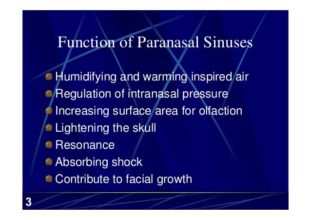
What is the main function of paranasal sinuses?
The function of the paranasal sinuses is a topic of much debate. Various roles have been suggested: Lightening the weight of the head; Supporting immune defence of the nasal cavity; Humidifying inspired air; Increasing resonance of the voice; The paranasal sinuses are formed during development by the nasal cavity eroding into the surrounding bones. All the sinuses therefore drain back into the nasal cavity – openings to the paranasal sinuses can be found on the roof and lateral nasal walls.
What are the dangers of a sinus infection?
- Fever
- Headaches
- Confusion and/or extreme drowsiness
- New-onset seizures
- Neck stiffness
- Vomiting
- Inattention
- Apathy and/or reduced motivation
- Personality changes, such as lack of emotional expression or inappropriate emotional responses
Where are the paranasal sinuses located?
There are three ethmoidal sinuses located within the ethmoid bone:
- Anterior – Opens onto the hiatus semilunaris (middle meatus)
- Middle – Opens onto the lateral wall of the middle meatus
- Posterior – Opens onto the lateral wall of the superior meatus
Is thickening of the mucosa dangerous?
Thickening of the mucosa of the paranasal sinuses is a common occurrence. It suggests mild sinusitis. If severe, sinusitis can cause frequent/vacuum headaches. It is a self-limiting and non-dangerous condition.
Are paranasal sinuses supposed to be aerated?
The paranasal sinuses are small, aerated areas within the bony skull that appear to provide several functions: They lighten the weight of the skull, produce a portion of upper airway respiratory mucus, and help with vocal resonance.
What is aerated paranasal sinuses?
The paranasal sinuses are aerated cavities within the skull that connect to the nasal cavity. There are four sets of paired sinuses: the maxillary, ethmoid, frontal, and sphenoid sinuses. The sinuses are lined with a pseudostratified, ciliated epithelium.
When are sinuses aerated?
Aeration of the ethmoid sinuses is first radiographically identified at 3 to 6 months of age. Posterior ethmoid air cell aeration is first visible around the age of 6 or 7 years. The sphenoid sinuses are tiny marrow-filled cavities at birth.
What is aerated mastoid air cells?
The mastoid air cells are thought to protect the delicate structures of the ear, regulate ear pressure and possibly protect the temporal bone during trauma. When the mastoid cells become infected or inflamed, often as a result of an unresolved middle ear infection (otitis media), mastoiditis can develop.
What is the meaning of well aerated?
well-aerated water. Definition English: Water aeration is the process of increasing the oxygen saturation of the water. ماء جيد التهوئة
How do you treat paranasal sinuses?
TreatmentNasal corticosteroids. ... Saline nasal irrigation, with nasal sprays or solutions, reduces drainage and rinses away irritants and allergies.Oral or injected corticosteroids. ... Allergy medications. ... Aspirin desensitization treatment, if you have reactions to aspirin that cause sinusitis and nasal polyps.More items...•
What does air-fluid levels in sinuses mean?
Air-fluid levels and complete opacification are more specific for sinusitis, but they are seen in only 60% of sinusitis cases. Air-fluid levels, as shown in the image below, generally indicate bacterial sinusitis. Air-fluid level (arrow) in the maxillary sinus suggests sinusitis.
Which paranasal sinuses are usually fully developed and aerated at birth?
The ethmoid air cells are usually fully developed and aerated at birth. True or False? Which paranasal sinuses are rarely symmetric in size and shape? Which sinuses may be absent (not developed)?
Which paranasal sinus is commonly infected?
Acute bacterial rhinosinusitis is inflammation of the paranasal sinuses, usually maxillary sinus, due to bacterial infection (Rosenfeld et al., 2015). The most common pathogens causing the disease are S. pneumoniae and H.
Does mastoiditis cause neck pain?
Mastoid infection may spread into the neck causing marked swelling on the side of the neck along with fever and exquisite tenderness. Infection in the neck left untreated can impair breathing and spread into the chest.
What is paranasal sinuses and mastoid air cells?
The paranasal sinuses of the craniofacial complex are air‐filled cavities, including the maxillary sinuses, the frontal sinuses, the sphenoid sinuses, and the ethmoid air cells.
Are mastoid air cells normal?
Concomitantly with growth, air cells develop in the normal mastoid by a process termed “pneumatization.” This process is governed by vital and anatomic factors, the influence of which causes each mastoid to attain an individual cell pattern which differs from that of its mate and is sufficiently characteristic for ...
What is excessive enlargement of the paranasal sinuses?
Excessive enlargement of the paranasal sinuses is a rare entity with an uncertain aetiology. In the medical literature, it has been described with many terms including hypersinus, pneumocele, pneumatocoele, sinus ectasia, hyperpneumatization, and pneumosinus dilatans. It usually affects the frontal sinus, although any sinus can be pathologically enlarged. We present a case of a patient with chronic headache, diagnosed with excessive aeration of all paranasal sinuses, together with atypical mastoid pneumatization. To the authors’ knowledge, this has not been previously reported in the literature.
Can a headache cause a enlarged sinus?
It usually affects the frontal sinus, although any sinus can be pathologically enlarged. We present a case of a patient with chronic headache, diagnosed with excessive aeration of all paranasal sinuses, together with atypical mastoid pneumatization.
Can you see the frontal sinus ostium endoscopically?
Although the frontal sinus os tium could not be visualized endoscopically on either side, no mucosal abnormalities were apparent at the area of the frontal sinus outflow tract. A high resolution CT scan revealed that all the paranasal sinuses and especially the frontal sinuses were excessively enlarged.
Is a headache a sinus disease?
It is widely accepted that headache is one of the symptoms of sinonasal diseases. The term “sinus headache” refers to a secondary headache associated with sinusitis that occurs when the sinuses become congested and obstructed because of paranasal sinus pathology.
Is surgery required for paranasal sinuses?
No surgical intervention was required for the patient , but in similar cases, with more severe symptoms, surgical treatment is a challenge for the surgeon and may mandate a multidisciplinary approach. 1. Introduction. Excessive enlargement of the paranasal sinuses is a rare entity with an uncertain aetiology.
Can mastoid pneumatization cause excessive aeration?
We present a rare case in the literature with excessive aeration of all paranasal sinuses, along with excessive mastoid pneumatization. Although no surgical intervention was required in this patient, in similar cases, with more severe symptoms, surgical treatment is a challenge for the surgeon and may mandate a multidisciplinary approach.
What is the first sinus in the paranasal sinuses?
The maxillary sinuses are the first of the paranasal sinuses to develop. In the neonate, the maxillary sinuses are quite small and may be partially or completely opacified. The maxillary sinuses grow progressively until the end of puberty. They reach the plane of the hard palate by 9 years of age. Asymmetry of the maxillary sinuses is common.
Which sinuses have the greatest degree of developmental variation?
The frontal sinuses have the greatest degree of developmental variation of any of the paranasal sinuses. Frontal sinus aeration can usually be identified on radiographs beginning at 4 to 8 years of age. The frontal, anterior ethmoid, and maxillary sinuses drain through the ostiomeatal complex (ostiomeatal unit).
When do sphenoid sinuses start to develop?
The sphenoid sinuses are tiny marrow-filled cavities at birth. Pneumatization usually begins around 2 years of age, and progresses until puberty. The mature sphenoid sinuses are bounded by the dura and sella superiorly, the cavernous sinuses laterally, posterior ethmoid air cells anteriorly, and the clivus posteriorly. The sphenoid sinuses are visible on radiographs beginning at 1 to 2 years of age.
When do maxillary sinuses become visible?
The maxillary sinuses become visible on standard radiographs at approximately 2 to 3 months of age. 1, 2. Small anterior ethmoid air cells are present at birth. There is rapid progression in ethmoid sinus development during the first 2 years of life. A second phase of rapid development occurs just before puberty.
Where does the posterior ethmoid air cell drain?
The posterior ethmoid air cells and the sphenoid sinus drain into the superior meatus, which is below the superior turbinate. The nasolacrimal duct drains into the inferior meatus. + +. Developmental variations of the nasopharynx and paranasal sinuses are common. Most are of no clinical significance.
Where is the drainage ostium located?
Asymmetry of the maxillary sinuses is common. The drainage ostium of the maxillary sinus is located superomedially. It empties into the infundibulum, which is located between the lamina papyracea and the uncinate process. The infundibulum opens into the middle meatus through the hiatus semilunaris.
Abstract
Introduction
- Excessive enlargement of the paranasal sinuses is a rare entity with an uncertain aetiology. In the medical literature, it has been described with many terms including hypersinus, pneumocele, pneumatocoele, sinus ectasia, hyperpneumatization, and pneumosinus dilatans. It usually affects the frontal sinus, although any sinus can be pathologically en...
Case Presentation
- A 38-year-old woman was referred for evaluation to the outpatient otorhinolaryngology clinic by the neurology department. The patient complained of intermittent episodes of moderate, nonthrobbing, and severe pressure-like headache since early adulthood. The headache was typically located at the anterior part of the cranium, mainly over the frontal, the anterior parietal, …
Discussion
- Terms like pneumosinus dilatans, pneumatocele, hyperpneumatization, sinus ectasia, sinus hypertrophy, and aerocele have been used in the literature to describe a hyperpneumatized paranasal sinus. An intuitive and simplified classification has been suggested by Urken et al. describing only three types of sinus hyperpneumatization [5]. Hypersinus refers to an enlargeme…
Conclusion
- We present a rare case in the literature with excessive aeration of all paranasal sinuses, along with excessive mastoid pneumatization. Although no surgical intervention was required in this patient, in similar cases, with more severe symptoms, surgical treatment is a challenge for the surgeon and may mandate a multidisciplinary approach.
Authors’ Contribution
- All the authors have made a significant contribution to the findings and methods in the paper. All the authors have read and approved the final draft.