What does apical Radiolucency mean? Background: Periapical radiolucency is the radiographic sign of inflammatory bone lesions around the apex of the tooth. Periapical radiolucency was defined as the presence of radiolucency or widening of the periapical periodontal ligament space to more than twice the normal width.
How to diagnose apical radiolucent lesion?
When an oral health professional is confronted with a case of apical radiolucent lesion, there is need to follow the laid down diagnostic technique before initiating therapy. This approach entails radiographic data to compile a diagnostic data base .
What is periapical radiolucency?
Periapical radiolucency is characterized by chronic or acute inflammatory lesions or lacerations around the apex of your tooth's root. It is usually triggered by bacterial invasion of the dental pulp and its presence is often an indication of poor oral health status.
What is the meaning of radiolucency?
ra·di·o·lu·cen·cy. Region of a radiograph showing increased exposure, either because of greater transradiancy of corresponding portion of subject or because of inhomogeneity in source of radiation, such as off-center positioning. radiolucency. the quality of being radiolucent.
What is a radiolucency area on an xray?
In areas having low-density, the x-ray beam passes through the structure easily, thus exposing/triggering the x-ray film/sensor. As a result, that portion of the picture appears darkened (referred to as radiolucent areas). When will a radiolucency (bone changes) show on an x-ray?
What causes apical radiolucency?
Most of periapical radiolucencies are the result of inflammation such as pulpal disease due to infection or trauma. Not all radiolucencies near the tooth root are due to infection. Odontogenic or non odontogenic lesion can over imposed with the apices of teeth.
What does radiolucency of a tooth mean?
It is common to see dark areas, known as radiolucencies, on a dental x-ray. A radiolucency often represents a void or an area of tissue that is less dense. Some of these radiolucencies are normal, such as those that represent openings in the jaw bone that allow certain nerves to enter and exit the jaw.
What does radiolucency mean on xray?
adjective Referring to a material or tissue that allows the facile passage of x-rays–ie, has an air or near air density; radiolucent structures are black or near black on conventional x-rays.
How is apical periodontitis treated?
How Do You Treat Apical Periodontitis?Root canal. In some cases, a root canal can minimize the inflammation of your gums by removing the bacteria and infected tissue from the tooth's pulp.Apicoectomy. If the infection develops or continues after the root canal, you might require an apicoectomy.
Can a root canal infection be seen on xray?
In the majority of cases, you will notice some type of symptom that indicates an infected root canal. Although, there are cases where there are no symptoms and the infection will only be found by your dentist on an x-ray.
What does radiolucency look like?
Radiopaque volumes of material have white appearance on radiographs, compared with the relatively darker appearance of radiolucent volumes. For example, on typical radiographs, bones look white or light gray (radiopaque), whereas muscle and skin look black or dark gray, being mostly invisible (radiolucent).
What is apical periodontitis?
Apical periodontitis is a chronic inflammatory disorder of periradicular tissues caused by aetiological agents of endodontic origin. Persistent apical periodontitis occurs when root canal treatment of apical periodontitis has not adequately eliminated intraradicular infection.
What is endodontic radiolucency?
In essence, the formation of an endodontic periapical radiolucency is evidence of the person's body creating a line of defense against the spread of bacteria and infection byproducts from within its associated tooth.
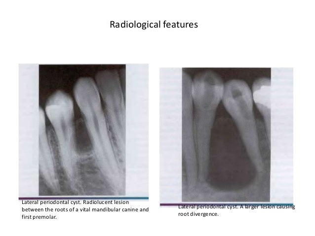
What is a radiolucent lesion?
The radiolucent lesion has a broad border of transition and has destroyed the lateral cortex of the bone. There is minimal reaction of the bone to the lesion. Another possible diagnosis is metastatic carcinoma.
Is apical periodontitis an infection?
Apical periodontitis is inflammation and destruction of periradicular tissues caused by etiological agents of endodontic origin. It is generally a sequel to endodontic infection (Fig. 1). Initially, the tooth pulp becomes infected and necrotic by an autogenous oral microflora.
What are the signs and symptoms associated with symptomatic apical periodontitis?
By far, most cases of apical periodontitis are asymptomatic. Pain, tenderness to biting pressure, percussion or palpation as well as swellings are typical clinical expres- sions of symptomatic apical periodontitis (Fig. 7.2a,b). The symptoms may vary from mild to severe.
Is apical periodontitis reversible?
Diagnosis: reversible pulpitis; normal apical tissues. If the pulp is exposed, treatment would be non-surgical endodontic treatment followed by a permanent restoration such as a crown.
What is periapical radiolucency?
A periapical radiolucency. To a dentist, this is proof positive that endodontic treatment is needed. Dentists refer to this type of dark spot as a “radiolucency.”. One centered on the tip of a tooth’s root (like in our picture) is referred to as a “periapical” radiolucency.
Why does radiolucency show up on x-rays?
As alluded to above, a radiolucency shows up on an x-ray because the bone in that region is less dense (it contains less mineral content, or else there is an actual void in the bone tissue in that area). When dental x-rays are taken: Areas having high-density show as white regions (referred to as radiopacities).
What is the dark spot on an x-ray?
a) Periapical radiolucencies. One of the most informative signs that shows up on an x-ray that indicates that a tooth possibly requires endodontic therapy is a dark spot that’s centered on the tip of the tooth’s root. A periapical radiolucency. To a dentist, this is proof positive that endodontic treatment is needed.
What is endodontic radiolucency?
In essence, the formation of an endodontic periapical radiolucency is evidence of the person’s body creating a line of defense against the spread of bacteria and infection byproducts from within its associated tooth. ▲ Section references – Hargreaves.
Why do x-rays show white?
That’s because the density of the object blocks the x-rays, and as a result that part of the x-ray film/sensor is shielded and remains unexposed, thus the object appears white or light in color.
Is a root canal diagnosis accurate?
So with that case, between the two (the deep cavity and the radiolucency at the tip of the tooth’s root, which equates with cause and effect), the dentist can feel essentially 100% confident that a diagnosis for root canal treatment is accurate. But other “dark spots” seen on films are much more difficult to interpret.
Can endodontic pathology be seen on x-rays?
It’s possible for a tooth to have advanced endodontic pathology but when an x-ray is taken of it everything about it and it’s surrounding tissues looks normal and healthy. This conundrum is easy enough to explain: The bone tissue changes that show up on a radiograph take time to develop (see below for further details).
What are periapical lucencies?
Periapical lucencies are often seen incidentally at head and neck imaging studies performed for indications not related to the teeth. These lesions are, however, occasionally manifestations of diseases that have a wide range of effects and may at times represent the source of symptoms that prompted the study. The vast majority of periapical lucencies are the result of apical periodontal or pulpal disease. If found in an advanced state or left untreated, disease related to the tooth may spread to adjacent tissues, including the sinuses, orbits, deep fascial spaces of the neck, and intracranial structures, and result in a significant increase in patient morbidity and mortality. Although the majority of periapical lucencies seen on radiographs and computed tomographic images occur secondary to apical periodontal or pulpal disease, not all lucencies near the tooth root are due to infection. Lucency near the tooth root may be seen in the setting of other diseases of odontogenic and non-odontogenic origin, including neoplasms. Although imaging findings for these lesions can include periapical lucent components, awareness of the varied secondary imaging features can aid the radiologist in developing an accurate differential diagnosis. Familiarity with the imaging features and differential diagnoses of diseases or conditions that cause lucency around the tooth root results in appropriate referral and prompt diagnosis, management, and treatment, and can prevent unnecessary additional imaging or intervention. In addition, early recognition and appropriate treatment of infectious processes will result in improved clinical outcomes and a decrease in morbidity and mortality.
What causes lucencies near the tooth root?
Nonapical periodontitis-related lucencies near the tooth root may arise from odontogenic cysts or tumors, including keratocystic odontogenic tumor (KCOT), dentigerous (follicular) cyst of the adjacent tooth, odontoma, and other odontogenic tumors such as ameloblastoma and odontogenic myxoma ( 5, 42, 43 ).
What causes periapical lesions?
According to one study, 78% of periapical lesions are the result of an infectious or inflammatory process, usually due to apical periodontal or pulpal disease ( 1 ). Recognition of the typical radiologic features of apical periodontal disease results in early referral and proper treatment.
Why is panoramic radiography used for maxilla?
Panoramic radiography can provide an overview of the maxilla and mandible and is frequently combined with intraoral radiography to increase the sensitivity and specificity of diagnosis of periapical lesions, because panoramic radiographs alone are often subject to variability in interpretation ( 9, 10 ).
Where is the pulp and root canal located?
The pulp and root canal are located within the center of the tooth and consist of the neurovascular bundle and connective tissue, both of which are radiolucent on radiographs and CT images. The neurovascular bundle enters the tooth via the apical foramen, located at the apex of the tooth root ( Fig 1a) ( 4, 6, 8 ).
Can apical periodontal disease cause sinusitis?
Apical periodontal disease of the maxillary teeth can extend into the maxillary sinuses, resulting in sinusitis. Odontogenic disease is thought to be responsible for 10%–12% of maxillary sinusitis cases ( 20 ). Patients with known apical periodontal disease have a twofold increased risk for developing maxillary sinusitis ( 26 ). Infection from an affected tooth can spread through the apical foramen into the periodontal ligament. Chronic infections and abscesses can result in disruption of the mucoperiosteum (Schneiderian membrane), creating a path of infection into the sinus. The apices of the second maxillary molars are the closest to the inferior maxillary sinus wall, with an average separation of only 1.7 mm ( 20 ).
What is a lucency in radiography?
What are lucencies? Radiographs, commonly known as X Rays, are images obtained for diagnostic purposes; in medical radiography, an X-ray generator produces a beam of energy (x-rays) that travels towards the body of the patient: part of these X rays will be absorbed by body structures while some of them will make it through ...
What does lucency mean in X-rays?
This means that X-rays were absorbed and didn’t make it through. A lucency is an area of low density, hence appearing black in color, often highlighted in the report because unexpected such as in a tissue that is supposed to be radiopaque (white in color).
What does a periodontal lucency mean?
In an OPG (orthopantomagram), an exam commonly prescribed by dentists, a periodontal or periapical lucency often indicates an infection. In a mammogram it may indicate a lipid cyst, a harmless lump of fatty tissue. In a bone radiograph, it may indicate an area of demineralization due to a trauma.
What does it mean when a radiograph is white?
A radiopaque structure has high density and will result in a white color on the radiograph. This means that X-rays were absorbed and didn’t make it through. A lucency is an area of low density, hence appearing black in color, often highlighted in the report because unexpected such as in a tissue that is supposed to be radiopaque (white in color).
What does CT scan show on a radiograph?
The obtained image on film will show radiolucent and radiopaque structures: A radiolucent structure has low density and will result in a black color on the radiograph. This means that X-rays made it through the body.
Introduction
A healthy 53-year-old male presented to our office on May 9, 2011, after being referred by a friend who was treated at our office. He was advised that he needed to have tooth No. 19 removed, and the extraction site grafted with bone and soft tissue.
Clinical and Radiographic Examination
patient had a 3-unit FPD on the left mandibular molar area extending from the second premolar, mesially, to the second molar (18-20) distally. A slight swelling was present, buccal to tooth No. 18, and the probing depths were surprisingly within normal limits, even when probed under anesthesia.
Medical history
Non-contributory. Patient was prescribed amoxicillin 500 mg TID for 2 days by his general dentist.
Treatment
Treatment was initiated with an I and D of the buccal swelling. The patient’s antibiotic regimen was changed to clindamycin 300 mg, sig 1 tab TID for 5 days. Retreatment followed by accessing the distal abutment of the 3-unit FPD. The cast post and existing gutta percha were removed.
Discussion
Management of teeth with previous root canal treatment that is failing requires more than just performing endodontic retreatment or surgery. The treating clinician must evaluate the cause of failure. These causes can range from being endodontic, restorative, periodontic, occlusion, patient’s habit (i.e. tongue ring), trauma, etc.