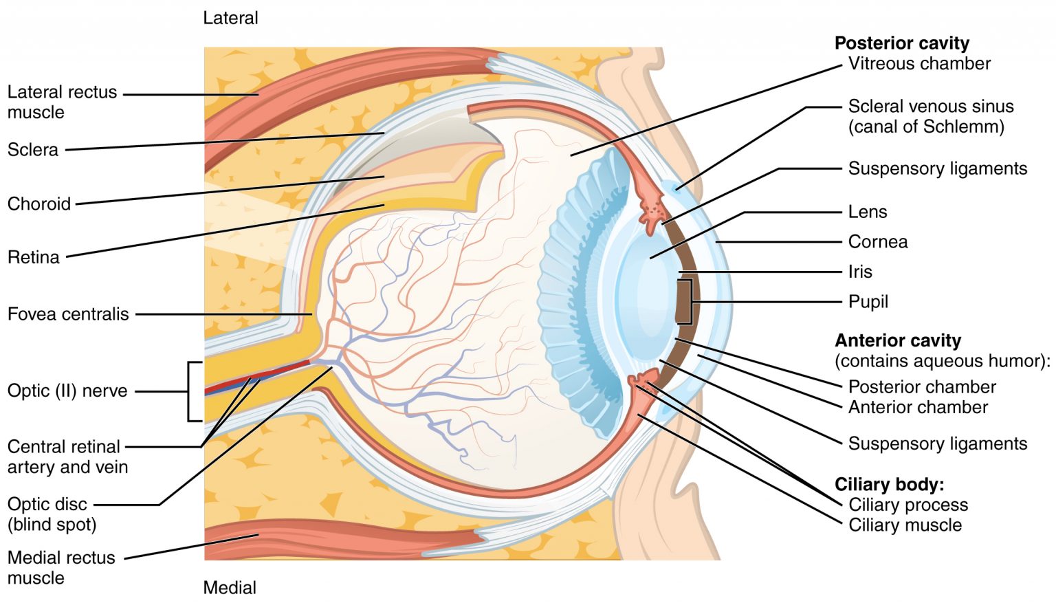
The Tunics of the Eye. From without inward the three tunics are: (1) A fibrous tunic, (Fig. 869) consisting of the sclera behind and the cornea in front; (2) a vascular pigmented tunic, comprising, from behind forward, the choroid
Choroid
The choroid, also known as the choroidea or choroid coat, is the vascular layer of the eye, containing connective tissues, and lying between the retina and the sclera. The human choroid is thickest at the far extreme rear of the eye, while in the outlying areas it narrows to 0.1 mm. The choroid provides oxygen and nourishment to the outer layers of the retina. Along with the ciliary body and iris, the choroid f…
What are the three layers of the eye?
know the three layers of the eye. fibrous tunic, vascular tunic, retina. function of fibrous tunic is located. external layer of the eye. the sclera and the cornea make up the following structure. limbus or corneal sclera junction.
What are the 3 tunics of the eye?
The Tunics of the Eye. From without inward the three tunics are: (1) A fibrous tunic, (Fig. 869) consisting of the sclera behind and the cornea in front; (2) a vascular pigmented tunic, comprising, from behind forward, the choroid, ciliary body, and iris; and (3) a nervous tunic, the retina.
What is the vascular tunic of the eye composed of?
the vascular tunic is composed from the following. choroid, the ciliary body and the iris. choroid. houses capillaries that supply nutrients and O2 to the retina and inner layer of the eye wall.
What are the three layers of the neural tunic of the eye wall quizlet?
What are the 3 layers tunics of the eye?
What are the 3 layers the wall of the eye is made up of?
What is neural tunic?
What makes up the neural tunic?
What are the 3 concentric layers of the eyeball mention the function of each layer?
What is the outer layer of the eye?
What are the three main parts of the eye?
The optical system of the human eye consists of three main components, i.e., the cornea, the crystalline lens and the iris. The iris controls the amount of light coming into the retina by regulating the diameter of the pupil.
Is the condition referred to as lazy eye?
Amblyopia, commonly known as lazy eye, is the eye condition noted by reduced vision not correctable by glasses or contact lenses and is not due to any eye disease. The brain, for some reason, does not fully acknowledge the images seen by the amblyopic eye.
What is the blind spot of the eye?
Blind spot, small portion of the visual field of each eye that corresponds to the position of the optic disk (also known as the optic nerve head) within the retina. There are no photoreceptors (i.e., rods or cones) in the optic disk, and, therefore, there is no image detection in this area.
What is the sclera made of?
The sclera, as separated from the cornea by the corneal limbus. The sclera, also known as the white of the eye, is the opaque, fibrous, protective, outer layer of the human eye containing mainly collagen and some elastic fiber.
How does the eye work?
Your eye works in a similar way to a camera. When you look at an object, light reflected from the object enters the eyes through the pupil and is focused through the optical components within the eye. The front of the eye is made of the cornea, iris, pupil and lens, and focuses the image onto the retina.
What is the function of the lens?
Lens. The lens is located in the eye. By changing its shape, the lens changes the focal distance of the eye. In other words, it focuses the light rays that pass through it (and onto the retina) in order to create clear images of objects that are positioned at various distances.
What carries nerve impulses from the eye to the brain?
The axons of the retina's ganglion cells collect in a bundle at the optic disc and emerge from the back of of the eye to form the optic nerve. The optic nerve is the pathway that carries the nerve impulses from each eye to the various structures in the brain that analyze these visual signals.
What are the three layers of the eye?
know the three layers of the eye. fibrous tunic, vascular tunic, retina. function of fibrous tunic is located. external layer of the eye. the sclera and the cornea make up the following structure. limbus or corneal sclera junction. corneas function. refract light coming into the cornea.
Which layer of the eye is vascular?
middle layer of the eye. the vascular tunic is composed from the following. choroid, the ciliary body and the iris. choroid. houses capillaries that supply nutrients and O2 to the retina and inner layer of the eye wall. melanocytes of choroid.
Which layer of the retina provides vitamin A?
layers of the retina. pigmented layer and neural layer. pigmented layer does the following. provides vitamin A for the photoreceptor cell and light rays that pass are absorbed. neural layer. houses the photoreceptors converting them into nerve impulses. orginization of the neural layer.
