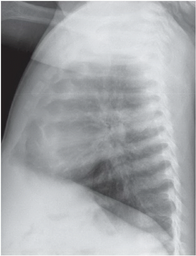
Costochondral calcification
- The anterior, medial ends of the ribs are normally cartilaginous, and usually are not visible on a chest X-ray
- With ageing the cartilage is increasingly calcified and can be particularly marked in some individuals, as in this image
What causes calcification of costal cartilage?
The pain associated with costochondritis usually:
- Occurs on the left side of your breastbone
- Is sharp, aching or pressure-like
- Affects more than one rib
- Worsens when you take a deep breath or cough
What is the function of calcified cartilage?
The zone of calcified cartilage (ZCC) forms an important interface between cartilage and bone for transmitting force, attaching cartilage to bone, and limiting diffusion from bone to the deeper layers of cartilage. The height of the ZCC is a relatively constant percent of articular cartilage and the height is maintained by a balance between progression of the tidemark into the unmineralized cartilage and changing into bone by vascular invasion and bony remodeling.
How do you cure costochondral separation?
What you can do
- Write down your symptoms, including any that may seem unrelated to the reason why you scheduled the appointment.
- Write down your key medical information, including other conditions and any history of injury to the painful joint.
- Write down key personal information, including any major changes or stressors in your life.
When does calcification of cartilage occur?
Calcification can be part of a normal healing response to musculoskeletal injuries. Calcifications are often found in arteries affected by arteriosclerosis (hardening of the arteries), in benign and malignant breast processes, at sites of bone or cartilage injury, and sometimes within cancers.
What causes costochondral calcification?
Early onset of costochondral calcification can be associated several endocrine and metabolic diseases, following a trauma, infections, malignancies or due to genetic factors and very rarely idiopathic. Our case exemplifies premature calcification of costal cartilages.
What causes rib calcification?
Be- fore that age, calcification of the costal car- tilages may be associated with chronic renal failure, thyroid disease, autoimmune disor- ders, and chondrosarcoma [2]. The normal patterns of calcification differ between males and females, usually appearing as peripheral parallel lines in males (Fig.
What is calcified costal cartilage?
Cartilage calcification occurs as calcium uptake into the cartilage tissue increases. The underlying mechanism for this difference between males and females in costal cartilage calcification patterns has not been fully explored.
What is costochondral?
Each of your ribs is connected to your breastbone by a piece of cartilage. The point where your rib connects to this cartilage is known as your costochondral joint. Costochondral separation is an injury that occurs when one or more of your ribs separates from this cartilage.
How do you treat calcification?
Most cases of calcific tendonitis can be treated with steroid injections, physical therapy and non-steroidal anti-inflammatory drugs (NSAIDs).
What are the symptoms of calcification?
Symptoms of calcificationBone pain.Bone spurs (occasionally visible as lumps under your skin)Breast mass or lump.Eye irritation or decreased vision.Impaired growth.Increased bone fractures.Muscle weakness or cramping.New deformities such as leg bowing or spine curvature.More items...
What causes a calcification?
Causes of calcification infections. calcium metabolism disorders that cause hypercalcemia (too much calcium in the blood) genetic or autoimmune disorders affecting the skeletal system and connective tissues. persistent inflammation.
Can costochondritis be seen on xray?
There is no laboratory or imaging test to confirm a diagnosis of costochondritis. But a health care provider might order certain tests, such as an electrocardiogram and chest X-ray, to rule out other conditions.
What happens to costal cartilage with age?
Overall, the costal cartilage midsubstance was slightly stiffer than articular cartilage, and did not show significant variation in stiffness with age or specimen calcification. Increased age did result in increased local variability of the indentation stiffness results.
Can Covid cause costochondritis?
Here, we report a case of severe costochondritis unresponsive to traditional management in a child who had COVID-19 infection a few months earlier. To our knowledge, this is the first reported case of post-COVID-19 costochondritis (PCC) that has been successfully managed with colchicine.
Is costochondritis life threatening?
Costochondritis is not life threatening and will not cause any other conditions. Medicine can help with the pain. These include acetaminophen (one brand: Tylenol), nonsteroidal anti-inflammatory drugs (such as aspirin or ibuprofen [one brand: Advil]), or other pain relievers, as appropriate.
Is costochondritis serious?
No, costochondritis isn't life-threatening. It's common for the chest pain to be misinterpreted as a heart attack. But costochondritis is not fatal. Treatments are available to help you heal from this condition.
What causes calcification of the costal cartilage?
What causes calcification of costal cartilage? There appears to be an association between heavy premature costal cartilage calc ification and certain systemic conditions, such as malignancy, autoimmune disorders, chronic renal failure, and thyroid disease, particularly Graves disease. Keeping this in consideration, ...
What is the role of costal cartilage?
Abstract Costal cartilage bridges the sternum and the ribs and plays a key role in the biomechanics of the chest. Costal cartilage is known to calcify in local regions with age, which can substantially stiffen its overall response to loading. Also Know, what happens to costal cartilage with age?
What is it called when a rib tears off?
When a rib tears away from the cartilage, the injury is called a costochondral separation. Similar Asks.
Where is costochondritis most commonly found?
Costochondritis. Costochondritis most commonly affects the upper ribs on the left-hand side of your body. Pain is often worst where the rib cartilage attaches to the breastbone (sternum), but it can also occur where the cartilage attaches to the rib.
What side of the breast does costochondritis occur?
The pain associated with costochondritis usually: Occurs on the left side of your breastbone. Is sharp, aching or pressure-like. Affects more than one rib. Worsens when you take a deep breath or cough.
What is the name of the pain in the chest wall?
Costochondritis is sometimes known as chest wall pain, costosternal syndrome or costosternal chondrodynia. Sometimes, swelling accompanies the pain (Tietze syndrome). Costochondritis usually has no apparent cause. Treatment focuses on easing your pain while you wait for the condition to improve on its own, which can take several weeks or longer.
How long does it take for Costochondritis to go away?
Costochondritis usually goes away on its own, although it might last for several weeks or longer. Treatment focuses on pain relief.
What is the pain in the chest called?
Costochondritis (kos-toe-kon-DRY-tis) is an inflammation of the cartilage that connects a rib to the breastbone (sternum). Pain caused by costochondritis might mimic that of a heart attack or other heart conditions. Costochondritis is sometimes known as chest wall pain, costosternal syndrome or costosternal chondrodynia.
What is a small staghorn calculus?
Renal stones can be small , resembling punctate foci of calcifications, or they may be large enough to fill the renal calyces and are known as staghorn calculi. The stones are also visible within the ureters, which travel down the lateral borders of the transverse processes of the lumbar spine.
What is a phlebolith?
Phleboliths are calcifications within the walls of the veins. They are common in the elderly and are often asymptomatic. Phleboliths are typically round, and have a lucent center and smooth borders.
What is abdominal radiograph?
Abdominal radiographs are part of routine diagnostic workup in patients presenting with an acute abdomen or abdominal trauma . They are obtained using conventional X-ray beams and represent the cornerstone of patient management before using computed tomography (CT).
Can gallbladder stones be seen on X-ray?
Gallbladder Stones. Gallbladder stones are mostly radiolucent, in contrast to renal tract stones, and are not visualized on X-ray. Only approximately 10% of gallbladder stones carry enough calcium to be radiopaque; therefore, an abdominal radiograph is not recommended for routine evaluation of gallstones.
Is a vascular calcification pathologic?
Arterial wall calcifications are usually pathologic in contrast to venous mural calcifications, which are usually benign. Arterial wall calcifications are strongly related to arterial stiffening and atherosclerosis. The points of vascular calcifications also correlate strongly with sites of arterial aneurysms.
Can a phlebolith be a ureteric stone?
Nonetheless, a phlebolith may be misdiagnosed as a ureteric stone warranting clinical evaluation. When in doubt, an abdominal CT scan should be ordered to clearly visualize the urinary system and accurately distinguish between renal tract calculi and phleboliths.