The axillary artery is a continuation of the subclavian artery that begins at the outer border of the first rib . It then courses through the axilla while being bordered by the lateral (superiorly), posterior (posteriorly), medial (inferiorly) cords of the brachial plexus and the ansa pectoralis (anteriorly).
Where is the axillary artery located?
The axillary artery, as a continuation of the subclavian artery, begins at the lateral margin of the first rib and ends normally at the inferior border of the teres major. The axillary artery supplies the walls of the axilla and related regions and continues as the brachial artery, to supply to more distal parts of the upper limb.
What is the function of the axillary artery?
Cutaneous innervation by the radial nerve is provided by the following nerve branches:
- Posterior cutaneous nerve of arm (originates in axilla)
- Inferior lateral cutaneous nerve of arm (originates in arm)
- Posterior cutaneous nerve of forearm (originates in arm)
What is mass of left axilla?
The lymph nodes of the axilla can be abnormal and symptomatic (presenting as an axillary mass or focal tenderness) with lots of conditions other than breast cancer. Some findings will be benign, such as infections – often related to infections in the tissues in the arm or the breast.
What is the anatomy of the ribs?
The stellate ganglion, which can be injured during central venous line placement is also known as the cervicothoracic ganglion. It is a large structure of up to 10 by 20 mm, formed by the fusion of the inferior cervical ganglion and the first thoracic ganglion.
Which of the following best demonstrates the right axillary ribs?
AP oblique rib projectionThe AP oblique rib projection is performed to best demonstrate the axillary ribs.
Which of the following positions can be demonstrated can be used to demonstrate the axillary ribs of the right thorax?
Thorax and AbdomenQuestionAnswerWhich of the following positions can be used to demonstrate the axillary ribs of the right thorax?LAO RPOWhich of the following positions will most effectively move the gallbladder away from the vertebrae in an asthenic patient?LAO168 more rows
Which position is best to visualize the upper ribs?
Sternum, ribs, sc joints by merrillsQuestionAnswerWhich position is best to visualize the upper ribs?Upright, seated or standingHow are the patients hands positioned on upright ribs?Hands against the hips with palms faced outwardsWhat are the breathing instructions for the upper ribs?Suspend at full inspiration53 more rows
How do you do oblique ribs?
4:006:03Adult Ribs-AP (Upper&Lower),AP Oblique(Upper&Lower) - YouTubeYouTubeStart of suggested clipEnd of suggested clipSo you'll find the mid sagittal. Point on the body in this lateral aspect and you go about halfway.MoreSo you'll find the mid sagittal. Point on the body in this lateral aspect and you go about halfway. In between that. So that would be our left oblique of the ribs.
Which projection best demonstrates the axillary portion of ribs?
AP oblique rib projectionThe AP oblique rib projection is performed to best demonstrate the axillary ribs. Oblique ribs may be conducted either as an anterior oblique or posterior oblique view.
What other position can be performed if the patient Cannot assume a prone position for the Rao sternum?
Questions and AnswersQuestionAnswerWhere is the central ray centered for the oblique and lateral projections of the sternum?Midsternum (midway between jugular notch and xiphoid.What other position can be performed if the patient cannot assume a prone position for the RAO sternum?LPO (oblique supine position)60 more rows
Which ribs are commonly fractured?
The most common ribs fractured are the 7th through 10th ribs. Fractures of the first and second ribs are rare but may be associated with serious damage to the brachial plexus of nerves, the subclavian vessels or associated with head, facial or thoracic aorta injuries.
What does a CT scan of the ribs show?
Doctors use chest CT to: examine abnormalities found on chest x-rays. help diagnose the causes of signs or symptoms of chest disease, such as cough, shortness of breath, chest pain, or fever. detect and evaluate the extent of tumors that arise in the chest, or tumors that have spread there from other parts of the body.
Does broken rib pain get worse before it gets better?
The worst pain is usually the first 1-2 weeks and gets gradually better after that. Healing time also depends on the person.
Are obliques ribs?
Origin. The internal obliques originate on the inguinal ligament, which is a ligament that runs from the anterior iliac spine to the pubic bone. Additionally they originate on the anterior iliac crest. The external obliques, however, originate on the lower eight ribs.
What is left posterior oblique position?
Left Posterior Oblique (LPO) Definition. The patient is lying recumbent or erect with left posterior surface against the IR, right side elevated; AP Oblique Projection. Term.
What is a chest PA and lateral?
0:000:33Normal Chest PA and Lateral - YouTubeYouTubeStart of suggested clipEnd of suggested clipThis is an example of a normal PA. And lateral chest x-ray in a patient who we imaged for a fever.MoreThis is an example of a normal PA. And lateral chest x-ray in a patient who we imaged for a fever. What you can see is that it's a fairly large patient.
Description
The axillary region (also known as the arm pit) is a pyramid-shaped area located between the shoulder girdle and thorax. It serves as a space for neurovascular and lymphatic structures to travel through to reach the upper extremity from the neck.
Clinical Relevance
Thoracic Outlet Syndrome (TOS) results from the compression of nerves and/or vessels at or around the apex of the axilla. The clinical presentation and distribution of TOS is dependent upon the structures compressed. A patient with TOS may complain of ipsilateral upper extremity pain, parasthesia, paresis, discoloration, and/or cold sensitivity.
Anatomy of the Human ribs
The Anatomy of the Human Ribs (costae) are one of the integral parts of the chest wall; they make up the lateral part of our body, its anterior and posterior wall and they entirely build the lateral parts of the chest wall.
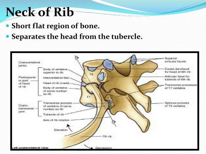
Parts of the Human Rib Bones
From the anatomy of the human rib cage, we can tell that the human Ribs bones have several parts: head (caput costae) neck (collum costae) body, corpus costae; and the front part of the rib. The head of the rib includes the articular surface, facies articularis capitis costae.
Properties of the ribs
A rib has a flat body, as you can see from the picture of the anatomy of the human rib cage. The upper edge is round and the lower sharp.
Course
The axillary artery is a continuation of the subclavian artery that begins at the outer border of the first rib .
Branches
The superior (highest) thoracic artery is the first branch of the axillary artery. It is given off proximal to the outer border of the anterior scalene muscle. It forms part of the arterial supply to the pectoral muscles .
Mnemonic
Here is a creative way to remember the branches of the axillary artery!
Sources
All content published on Kenhub is reviewed by medical and anatomy experts. The information we provide is grounded on academic literature and peer-reviewed research. Kenhub does not provide medical advice. You can learn more about our content creation and review standards by reading our content quality guidelines.
Overview
A fractured rib occurs when one of the bones in your rib cage breaks or cracks.
Symptoms
The pain associated with a broken rib usually occurs or worsens when you:
Causes
Broken ribs are most commonly caused by direct impacts — such as those from motor vehicle accidents, falls, child abuse or contact sports. Ribs also can be fractured by repetitive trauma from sports like golf and rowing or from severe and prolonged coughing.
Complications
A broken rib can injure blood vessels and internal organs. The risk increases with the number of broken ribs. Complications vary depending on which ribs break. Possible complications include:
Description
- The axillary region (also known as the arm pit) is a pyramid-shaped area located between the shoulder girdle and thorax. It serves as a space for neurovascular and lymphatic structures to travel through to reach the upper extremity from the neck.
Structure/Borders
- Apex: (also known as cervicoaxillary margin, axillary inlet) region of entrance/exit of the axillary neurovascular bundle
- Base:
- Anterior Wall:
- Posterior Wall:
Contents
- The axillary artery is an extension of the subclavian artery, and is called so after passing the first rib. It is renamed and considered the brachial artery after passing the teres major and exiting the axilla. The artery is encompassed by the axillary sheath and the brachial plexus cords and branches. The axillary artery is divided into three part...
Clinical Relevance
- Thoracic Outlet Syndrome (TOS) results from the compression of nerves and/or vessels at or around the apex of the axilla. The clinical presentation and distribution of TOS is dependent upon the str...
Related Pages