What is the nictitating membrane?
The nictitating membrane is a semilunar fold of conjunctiva, which also occurs in redimentary form in humans and non-human primates, that protrudes from the medial canthus and can extend over a significant portion of the cornea.
Do any animals have a nictitating membrane?
Some mammals, such as cats, camels, polar bears, seals and aardvarks, have full nictitating membranes. Often called a third eyelid or haw, it may be referred to in scientific terminology as the plica semilunaris, membrana nictitans, or palpebra tertia.
Why did the nictitating membrane disappear in humans?
Nictitating Membrane. In some species, the membrane is sufficiently transparent so as to enable vision when underground or underwater. Though the reason for the loss of a nictitating membrane in humans in unclear, changes in habitat and eye physiology may have rendered the tissue unnecessary.
What does the nictitating membrane do in a chimpanzee?
Nictitating Membrane. In the chimpanzee, however—one of the human species’ closest relatives—the plica semilunaris also appears to be vestigial. The function of the nictitating membrane in many animals is protective—for example, keeping the eye clean and moist or concealing the iris from predators.
Why do humans not have a nictitating membrane?
In some species, the membrane is sufficiently transparent so as to enable vision when underground or underwater. Though the reason for the loss of a nictitating membrane in humans in unclear, changes in habitat and eye physiology may have rendered the tissue unnecessary.
Did humans have 3rd eyelids?
It's actually the remnant of a third eyelid. In humans, it's vestigial, meaning it no longer serves its original purpose. There are several other vestigial structures in the human body, quietly riding along from one of our ancestor species to the next.
Is nictitating membrane vestigial in humans?
In humans, this membrane is also called the semilunar fold. Hence the nictitating membrane is vestigial in humans, so the correct answer is option C.
What is the function of nictitating membrane in humans?
The nictitating membrane (from Latin nictare, to blink) is a transparent or translucent third eyelid present in some animals that can be drawn across the eye from the medial canthus to protect and moisten it while maintaining vision.
Why do I have 4 eyelids?
In most cases, an extra eyelid crease is caused by: loss of skin elasticity and weakened connections between the skin and muscle beneath. soft tissue thinning and loss of fat under the skin in the upper eyelid, above your natural eyelid crease.
Did humans have a tail?
Many believe that human ancestors had and used some form of a tail. Over time as a species, however, we evolved past the need for such an organ, which is why the majority of humans no longer grow them. Most humans grow a tail in the womb, which disappears by eight weeks.
How humans lost their tails?
Recently, researchers uncovered a genetic clue about why humans have no tails. They identified a so-called jumping gene related to tail growth that may have leaped into a different location in the genome of a primate species millions of years ago. And in doing so, it created a mutation that took our tails away.
Why do dogs have a 3rd eyelid?
These third eyelids serve four purposes: Protect the eye from injury. Keep the cornea clean. Act as a lymph node which produces antibodies to protect against infection.
Why do humans have a tailbone but no tail?
The tail vanishes by the time humans are born, and the remaining vertebrae merge to form the coccyx, or tailbone. Tailbones helped our ancestors with mobility and balance, but the tail shrank as humans learned to walk upright. The coccyx now serves no purpose in humans.
Do mammals have nictitating membrane?
The nictitating membrane is a transparent or translucent third eyelid present in some animals that can be drawn across the eye for protection and to moisten it while maintaining visibility. Fully developed nictitating membranes are found in fish, amphibians, reptiles, birds, and mammals but are rare in primates.
Can you have a triple eyelid?
Triple eyelid occurs when the upper eyelid has two folds instead of one. Several factors may be involved, but in most cases it is caused by redundant skin, fat atrophy, or an improper functioning of the eyelid's fibrous muscle tissue.
Where is the nictitating membrane found?
The nictitating membrane (third eyelid, nictitans) is located ventromedially between the lacrimal caruncle and the globe.
Where is the nictitating membrane located?
Nictitating membrane (plica semilunaris conjunctivae, third eyelid) In dogs and cats the nictitating membrane is located nasoventrally, in the rabbit nasally , and in birds dorsally. In most birds the third eyelid is transparent.
What is the function of the nictitating membrane?
The nictitating membrane, or third eyelid, serves to protect the globe, distribute tears, and produce immunoglobulins and part of the precorneal tear film. The caruncle is a small, finely haired prominence within the nasal canthus that may or may not be pigmented. The nictitating membrane has an exposed, palpebral conjunctival surface ...
What does scraping of the bulbar surface of the membrane in normal or diseased eyes resemble?
Scrapings of the bulbar surface of the membrane in normal or diseased eyes may resemble cytological preparations from lymph nodes with all expected types of lymphoid cells. As a conjunctival surface, the nictitating membrane may be affected by most of the diseases of the conjunctiva described previously.
Why do horses have nictitating flaps?
Adaptations in large animals and special species. Nictitating membrane flaps provide more support to the diseased cornea than the temporary complete tarsorrhaphy in horses. Nictitating membrane flaps are used to cover and protect a weakened cornea, but are not usually a source of tissues for the cornea.
Which membrane is in contact with the globe?
The nictitating membrane has an exposed, palpebral conjunctival surface and a bulbar conjunctival surface that is in contact with the underlying globe. Superficial lymphoid follicles are located on the bulbar conjunctival surface and presumptively produce IgA, which becomes a component of the tear film.
How do nititating flaps work?
Nictitating membrane flaps provide more support to a diseased cornea than a temporary complete tarsorrhaphy. They are used to cover and protect a weakened cornea , but are not a source of nutrients or collagen to replace corneal tissue loss. Nictitating membrane flaps are recommended for superficial corneal diseases, as well as to reinforce a bulbar conjunctival graft. Third eyelid flaps are contraindicated for most corneal ulcers in horses, especially the melting ulcers that progress rapidly because they do not provide a blood supply or fibrovascular tissues to the ulcer. In addition, it is impossible to visually observe the progression of the disease and cover the normal cornea. Furthermore, third eyelid flaps might impede the penetration of topical medication and retain inflammatory exudates adjacent to the lesion. To place a third eyelid flap, one tip of the forceps (Bishop–Harmon forceps) is placed above and the other tip below the lateral aspect of the upper eyelid margin. The forceps tips are inserted as far as possible into the conjunctival fornix where the sutures should traverse the eyelid. The first suture (4-0 non-absorbable suture) with a pre-placed stent (intravenous tubing, buttons, polystyrene foam strip, rubber bands) is passed through the eyelid skin and into the conjunctival fornix. The suture is then passed through the palpebral (anterior) side of the nictitating membrane to encircle the upper stem of the T-shaped nictitating hyaline cartilage. The suture is then finally passed through the dorsolateral conjunctival fornix, the upper eyelid, and the stent again. The nictitating membrane is then protracted until its leading margin is at the limbus or within the conjunctival fornix.
What is the nictitating membrane?
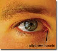
Nictitating Membrane. The plica semilunaris is a fold of conjunctiva at the inner corner of the human eye. Its likeness to the nictitating membrane, or third eyelid, of other animals led to the idea that it might be the vestige of such a structure, which is still part of the eye in some primates, including gorillas.
Why is the nictitating membrane transparent?
In some species, the membrane is sufficiently transparent so as to enable vision when underground or underwater. Though the reason for the loss of a nictitating membrane in humans in unclear, changes in habitat and eye physiology may have rendered the tissue unnecessary.
Can a human have a tail?
On rare occasion, a human infant is born with a vestigial tail. In modern medical literature, such tails lack vertebrae and typically are harmless, though some are associated with spina bifida (failure of the vertebrae to completely enclose the spinal cord). Tails in human infants typically are removed through surgery without complication.
Overview
Distribution
Fully developed nictitating membranes are found in fish, amphibians, reptiles, birds and mammals, but are rare in primates. In humans, the plica semilunaris (also known as the semilunar fold) and its associated muscles are homologous to the nictitating membranes seen in some other mammals and other vertebrates. In most primate species, a plica semilunaris is generally not prese…
Description
The nictitating membrane is a transparent or translucent third eyelid present in some animals that can be drawn across the eye for protection and to moisten it while maintaining vision. The term comes from the Latin word nictare, meaning "to blink". It is often called a third eyelid or haw, and may be referred to in scientific terminology as the plica semilunaris, membrana nictitans, or palpebr…
Functions
The nictitating membrane is normally translucent. In some diving animals, including sea lions, it is activated on land, to remove sand and other debris—its function in most animals. In crocodiles, it protects their eyes from water but also hinders their focus under water. In some diving animals, for example beavers and manatees, it is transparent and moves across the eye to protect it while under …
Vestigiality
Nictitating membranes in cats and dogs do not have many muscle fibers, so they are not usually visible; chronic visibility should be taken as a sign of poor condition or ill health. The membrane can, however, be seen clearly when gently opening the eye of the healthy animal when it is asleep, or by pushing down/applying pressure on the eyeball, which will cause it to appear. In some …
See also
• Accessory abducens nucleus
• Human vestigiality
External links
• Hawk, transparent eyelids (nicitating membrane slow motion video) on YouTube